
Monday, October 29, 2007 - 11:38 AM
13288
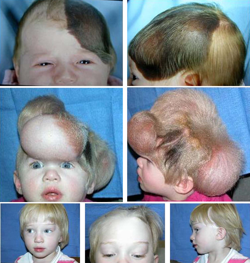
Pediatric Tissue Expansion for Forehead Reconstruction: A 13-Year Review and an Algorithm for its Use
Purpose: Reconstruction of the forehead in children when 25% or more of the forehead is involved presents a complex reconstructive challenge due to the confluence of highly visible aesthetic units. Tissue expansion of adjacent uninvolved tissues provides the best option for forehead reconstruction, providing like tissues without displacing key anatomic landmarks. The present study was performed to develop an algorithm that will help to optimize the outcome of pediatric forehead reconstruction for lesions involving 25% or more of the forehead.
Methods: A 13-year retrospective review was performed of all pediatric patients who completed reconstruction for lesions involving at least 25% of the forehead by a single surgeon (AKG). All lesions were classified based on percent of forehead involved and involvement of adjacent tissues (scalp, cheek, and eyebrow).
Results: Twenty patients completed reconstruction for lesions involving 25% or more of the forehead, all of whom had tissue expansion. Patients ranged in age from 9 months to 10 years at the time of the initial expansion. Seventeen patients presented with giant congenital pigmented nevi, and one patient each presented with nevus sebaceous, post-traumatic scarring, and post-involutional hemangioma. The median number of surgical procedures required to complete forehead reconstruction was 6 (range 2-11), involving a median of 3 tissue expansion procedures (range 1-4). Simultaneous expanders were placed in the scalp (16 patients) and in the cheek (8 patients). Eight patients required additional serial excision to complete forehead reconstruction. Five patients underwent correction of eyebrow ptosis at a final procedure. Reconstruction involved 25% to 65% of the forehead in 19 patients, 17 of whom were reconstructed with serial forehead expansion and advancement flaps (Figure). One patient presented with a pigmented nevus occupying over 75% of the forehead. In this patient there was not adequate adjacent tissue to reconstruct the forehead with expanded advancement flaps, and reconstruction was performed with an expanded full-thickness skin graft from the lower abdomen. The entire extent of the visible lesion was excised and complete skin coverage achieved in all patients.
Conclusions: Reconstruction of 25% or more of the forehead in children is best accomplished using tissue expansion. Up to 65% of the forehead can be reconstructed through expansion and direct advancement of adjacent tissues. Simultaneous expansion should be done in the cheek for lesions which extend below the lateral canthus, and in the scalp for lesions that extend beyond the hairline. Correction of brow ptosis is required in 25% of cases, and should be reserved for the final procedure. Lesions requiring reconstruction of more than 65% of the forehead are best accomplished with distant tissues. The latter could be performed with an expanded full-thickness skin graft to resurface the entire forehead as one aesthetic unit, or with free tissue transfer. Through a carefully planned application of tissue expansion to pediatric forehead reconstruction, all defects can be reconstructed reliably and by aesthetically accepted principles.
See more of Cranio/Maxillofacial/Head & Neck
Back to 2007am Complete Scientific Program