
Sunday, October 3, 2010 - 10:10 AM
17443
Utilizing Reverse End-to-Side Neurorrhaphies in Peripheral Nerve Injuries
Poor functional recovery following repair of a peripheral nerve injury is proportional to the time and inversely proportional to the number of motor axons able to reestablish their synaptic connections1. Motor-sensory preservation utilizes local nerve transfers to support denervated muscle, by providing it with early temporary innervation, and to supply additional motor axons to augment recovery in the event that the original motor axons incompletely regenerate to their targets. This concept of motor-sensory preservation can be utilized through reverse end-to-side neurorrhaphies and can be employed as a technique to manage peripheral nerve injuries.
36 Lewis rats were divided into 3 groups (Fig 1.). In Group 1, the right tibial nerve is transected.Ā The proximal stump is doubly ligated, sewn into a blind ended silicone cap, and buried within muscle. In Group 2, theĀ peroneal nerve is transected distally and the tibial nerve is transected 2.5mm proximal to the first branch. The proximal tibial nerve stump is treated as described in Group 1.Ā The proximal peroneal nerve stump is coapted to the distal tibial nerve in in an end-to-end fashion. In Group 3, the tibial nerve is transected approximately 4.5mm proximal to the first branch. A perineurial window is created, with care to not damage underlying endoneurial tubes, into the side of the tibial nerve approximately 2.5mm proximal to the first branch (distal to transection site). The peroneal nerve transection and proximal tibial nerve stump treatment is described in Group 2. The proximal peroneal nerve stump is coapted to the perineurial window in the tibial nerve in a reverse end-to-side fashion. Evaluated outcomes include muscle mass, motor end plate counts, and nerve histomorphometry. 5 male transgenic GFP Sprague Dawley rats, expressing GFP in neural tissue, underwent the same procedure as described for Group 3. In these animals, axonal regeneration is visualized over time using confocal microscopy for qualitative assessment (Fig 2.).
Rats undergoing reverse end-to-side neurorrhaphy showed improved muscle mass and higher motor end plate and nerve fiber counts. Confocal microscopy demonstrates axon regeneration through the reverse end-to-side coaptation into the recipient nerve.
1.ĀĀĀĀĀĀĀĀĀĀĀ Lien SC, Cederna PS, Kuzon WM, Jr.: Optimizing skeletal muscle reinnervation with nerve transfer. Hand Clin 24:445-454, vii, 2008
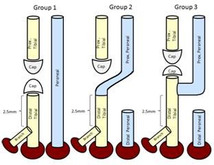
Fig 1. Schematic Diagram of Groups
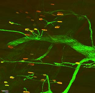
Fig 2. Confocal Imaging of GFP Transgenic Rat Nerve
See more of Hand & Upper Extremity (5 mins)
Back to 2010am Complete Scientific Program