
Monday, November 4, 2002 - 2:51 PM
784
Closure of Palatial Fistulas Using Acellular Dermal Grafts
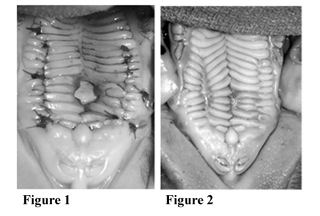
Purpose: Palatal fistulas are a common complication of palatoplasty. Surgical repair is indicated to eliminate associated problems, including nasal leakage of food and fluids, nasal air emission, and chronic inflammation. The presence of scar tissue and deficiency of healthy local tissues make tension-free closure of palatal fistulae difficult, contributing to failure rates of 10% to 65% and often necessitating the use of regional flaps. This study examines the efficacy of a novel technique for palatal fistula repair using acellular dermal grafts for simple, tension-free closure. Methods: 10mm oronasal fistulae were created in the hard palate of 3-week-old Yorkshire piglets. The bony palate was ronguered 2mm beyond the mucosal edges to allow oral-to-nasal re-mucosalization. In one group of animals (n=3), the fistulae were allowed to heal for 2 weeks prior to repair. In each animal, bilateral mucosal incisions were made, and the mucoperiosteum was elevated from the underlying bone. Acellular dermal grafts were then inset beneath the oral mucosa, traversing the palatal fistula. No attempts were made to suture the mucosal edges of the fistulae (Figure 1). Three weeks postoperatively, animals were sacrificed and the palates harvested for histologic examination. The fistulae of a second group of animals (n=3) remained unrepaired and were examined five weeks after their creation. Results: All animals demonstrated patent oronasal fistulas with complete re-mucosalization of the wound margins at two weeks. Those animals that underwent fistula repair with the acellular dermal grafts demonstrated complete healing at three weeks (Figure 2), with complete re-epithelialization and cellular infiltration into the grafts. Unrepaired fistulae remained patent at five weeks. Conclusion: This study demonstrates the successful closure of palatal fistulas in an animal model using interposition grafts of acellular dermis. This novel method offers promise as a simple and effective technique for tension-free closure of oronasal fistulas after primary palatoplasty.
See more of Cranio/Maxillofacial (Including Facial Fractures, Cleft-Lip and Palate)
Back to 2002 Complete Scientific Program
Back to 2002 Meeting home