Sunday, November 3, 2002
242
Galea-Frontalis Flap in Frontal Sinus Fracture Management
Introduction: Frontal sinus fracture management is still controversial and involves preserving function when feasible or obliterating the sinus and duct depending on the fracture pattern. There is no single algorithm for the choice of cranialization versus obliteration as well as the method of obliteration. In this study during initial treatment of frontal sinus fractures or for their late complications an anteriorly based galea frontalis myofascial flap is used.
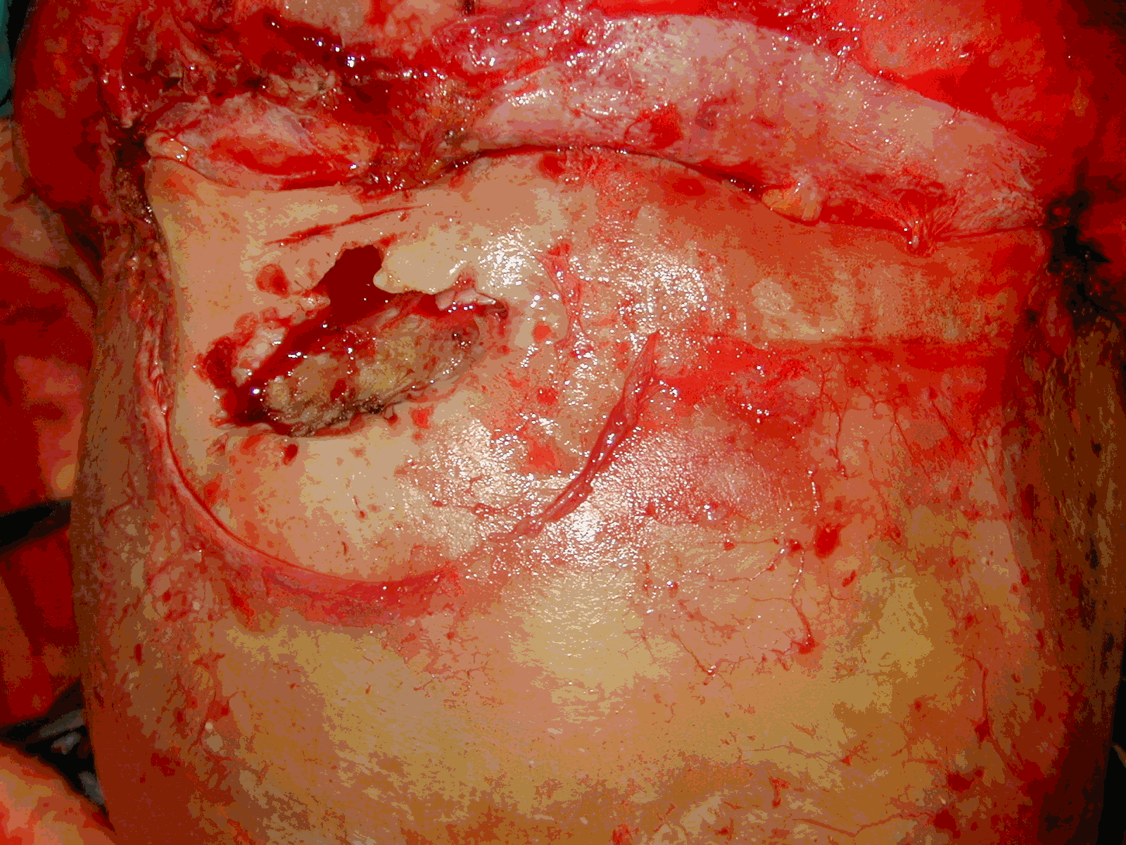
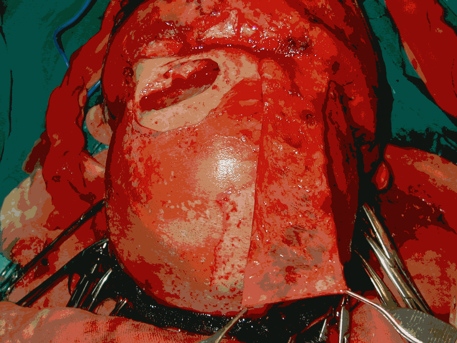
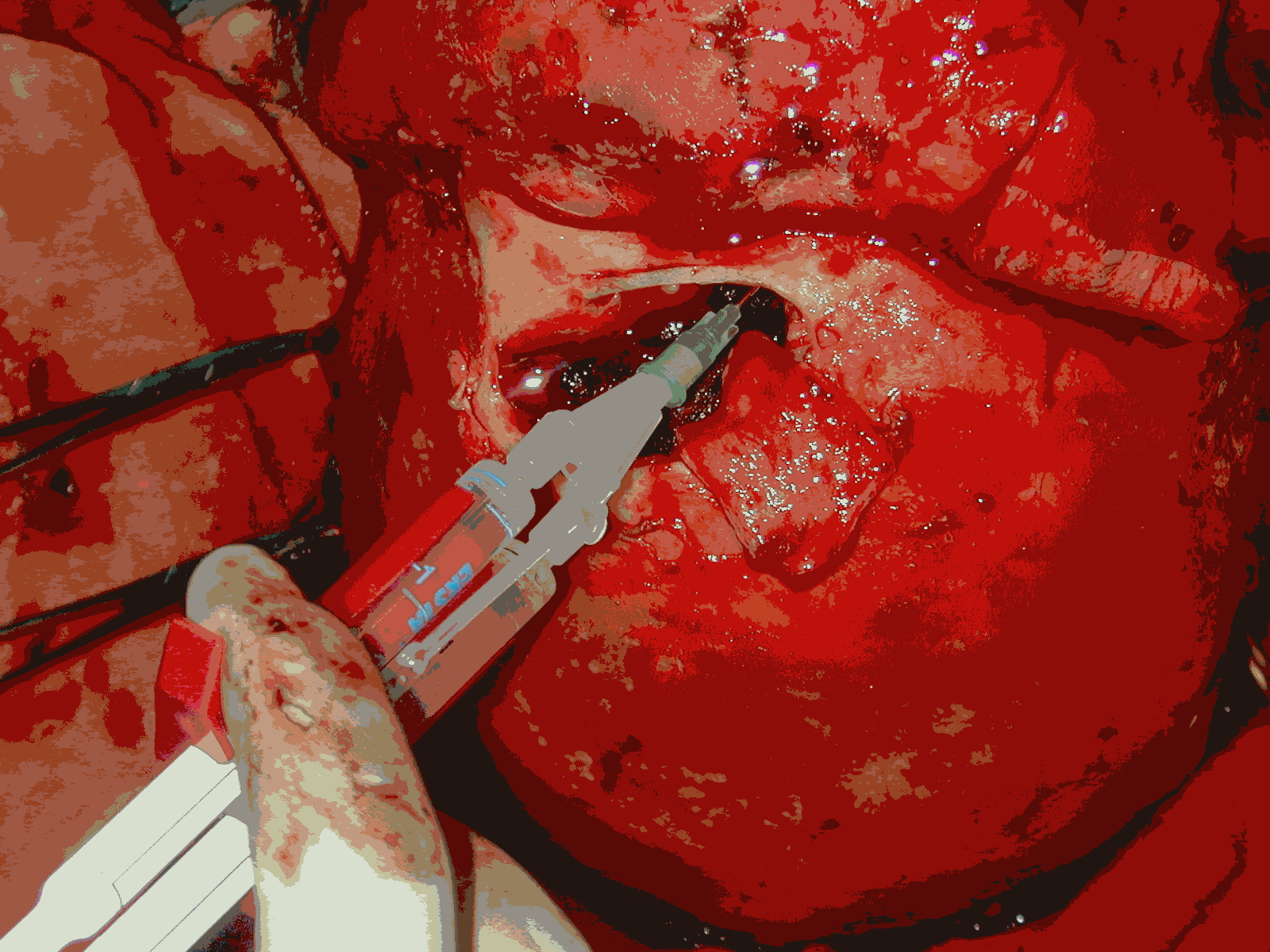
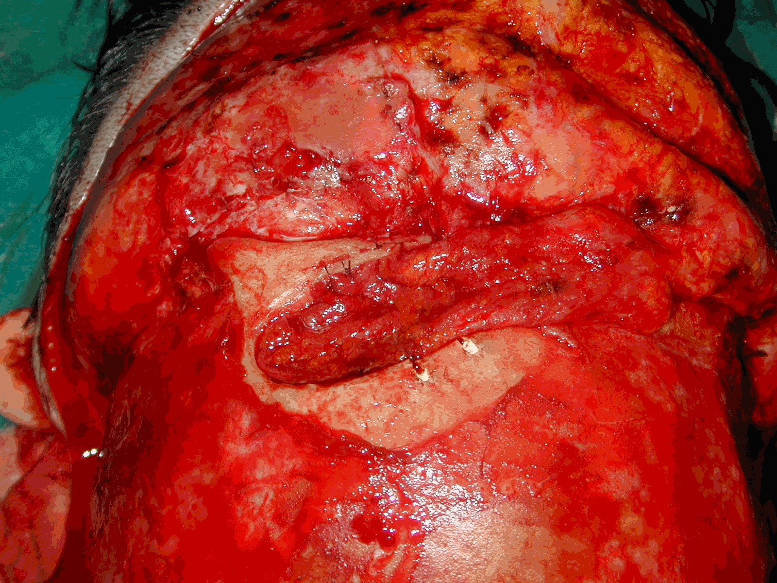
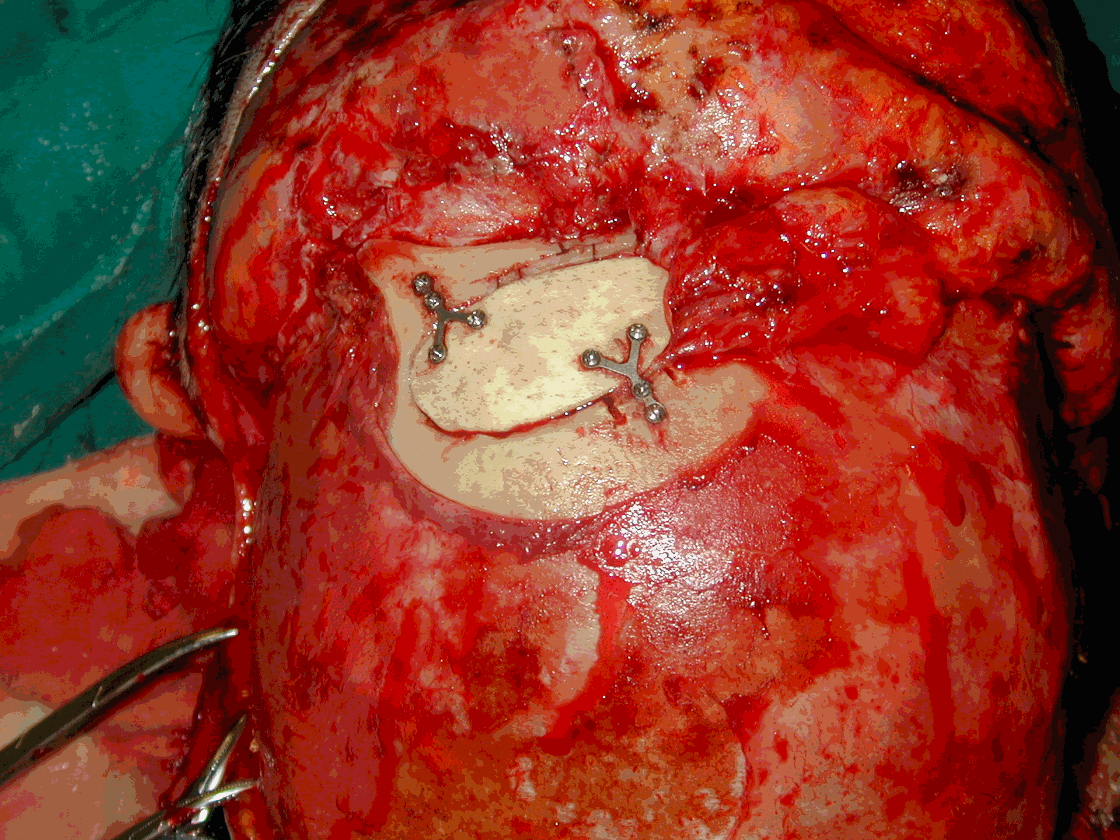
Materials and Methods: Twelve patients with frontal sinus fractures involving either the posterior wall and/or nasofrontal duct have been included into the study. Through a bicoronal approach hard tissue reconstruction and dural repair if required were performed. To obliterate the frontal sinus a flap is elevated from posterior to anterior including the galea and frontalis muscle. This flap is brought into the sinus and fixed in place by using both sutures and fibrin glue following mucosal removal. Then the anterior wall defect is reconstructed with cranial bone grafts (figure).
Results: Follow up ranges from 8 to 36 months during which none of the patients have experienced any early or late complication. In four patients with dural tear, no postoperative cerebrospinal fluid escape was observed. The elevation of the flap caused a partial sensory loss over the forehead skin, which usually resolved over a period of six months with no other additional donor site morbidity.
Conclusion: The anteriorly based galea frontalis myofascial flap is a reliable tissue with its blood supply mainly coming from supraorbital vessels. Two flaps can be elevated from each side when both sides of the sinus need to be obliterated. Especially following a dural repair, instead of bringing a non vital tissue for obliteration, this flap provides a more secure barrier. With these properties anteriorly based galea frontalis flap seems to be one of the best options for sinus obliteration as it provides a vascularised tissue to cover the anterior cranial base.
View Synopsis (.doc format, 149.0 kb)
See more of Cranio/Maxillofacial (Including Facial Fractures, Cleft-Lip and Palate)
Back to 2002 Complete Scientific Program
Back to 2002 Meeting home
