Wednesday, October 29, 2003
3559
P86: Use of External Skin Expansion in the Treatment of Benign and Malignant Lesions of the Face
Purpose:
To demonstrate the use of external skin expansion in treating large benign and malignant lesions of the face without causing distortion.
Material:
External skin expander (Photo #1). External expander creates a constant continuous pressure of 460 grams. They consist of two pins and a reciprocal spring. Pins are placed approximately 2-3 cm from skin edge. These work best under tension.
Method:
The technique involves a combination of vigorous wound toilette, the judicious use of sutures and tissue expansion produced by the application of specially designed external tissue expanders. Gradual approximation of the wound edges is achieved with final by suture.
Basic Principle:
1) Approximation (as much as possible) of wound edge with 2o nylon.
2) Application of expander. Allow expander to stretch skin and subcutaneous tissue daily.
3) Remove expanders, sterilize expanders in 50% Alcohol and 50% Betadine solution for 20 minutes.
4) Approximation of tissue with 2o nylon.
5) Re-application of expanders
6) When adequate tissue is available, freshen edges and close wound.
Case study
Pre-expansion of benign lesion.
Case #1 Nevi of face
23 year old female with congenital nevi on left side of face and nose.
Preoperative face and nose - Case 1 Photo 1
Application of expanders - Case 1 Photo 2
Dressing combine King and Unna boot - Case 1 Photo 3
6th day (4 days daily expansion) - Case 1 Photo 4
6 months follow up - Case 1 Photo 5
1 year follow up - Case 1 Photo 6
Case #2
32 year old male with nevi of forehead.
Preoperative photo - Case 2 Photo 1
3 days expansion and excision - Case 2 Photo 2
1 day post excision expansion - Case 2 Photo 3
11 days post excision - Case 2 Photo 4
2 months follow up - Case 2 Photo 5
9 months follow up - Case 2 Photo 6
B) Pre-expansion of a malignant lesion.
Case #3
62-year-old male with enlarged ulcerating squamous cancer cell of lower lip.
Preoperative lesion - Case 3 Photo 1
Preoperative marking - Case 3 Photo 2
Preoperative approximation - Case 3 Photo 3
External skin expansion - Case 3 Photo 4
Excision of lesion of lower lip - Case 3 Photo 5
Postoperative expansion - Case 3 Photo 6
6 days postoperative - Case 3 Photo 7
5 month follow up - Case 3 Photo 8-9
C) Intra operative expansion of a malignant lesion.
Case #4
79 year old male with basal cell cancer of nose and left pre-auricular area.
Preoperative lesions - Case 4 Photo 1
Intra operative expansion while excising- Case 4 Photo 2
and grafting nasal lesion
Excision of lesion of pre-auricular area - Case 4 Photo 3
7th postoperative day - Case 4 Photo 4
7 months follow up - Case 4 Photo 5
Total number of cases:
16 benign lesions
15 malignant lesions
Conclusion:
The case of preoperative and intra-operative expansion of skin greatly facilitates the reconstruction of large benign and malignant tumor without extensive undermining or distortion.
Photos Photos Case 1
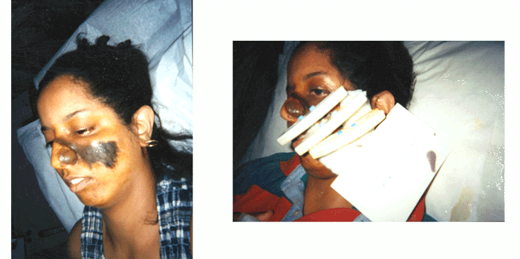
Photo 1 Photo 2
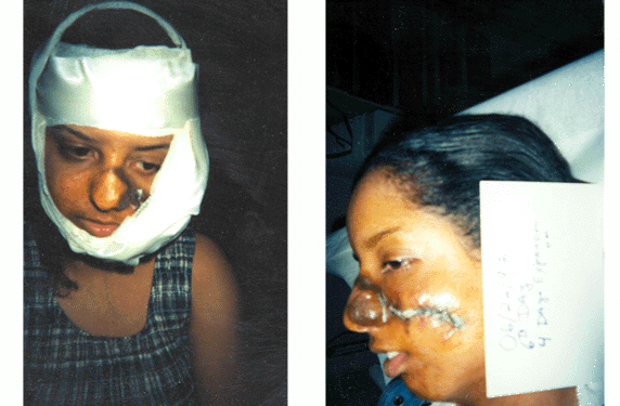
Photo 3 Photo 4

Photo 5 Photo 6 Photos Case 2
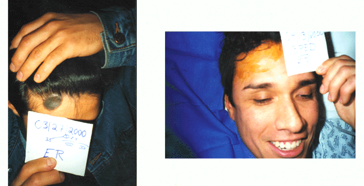
Photo 1 Photo 2
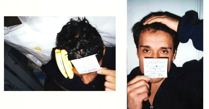
Photo 3 Photo 4
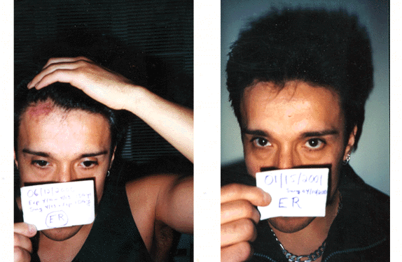
Photo 5 Photo 6 Photos Case 3
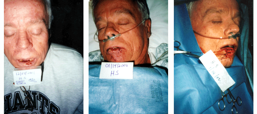
Photo 1 Photo 2 Photo 3
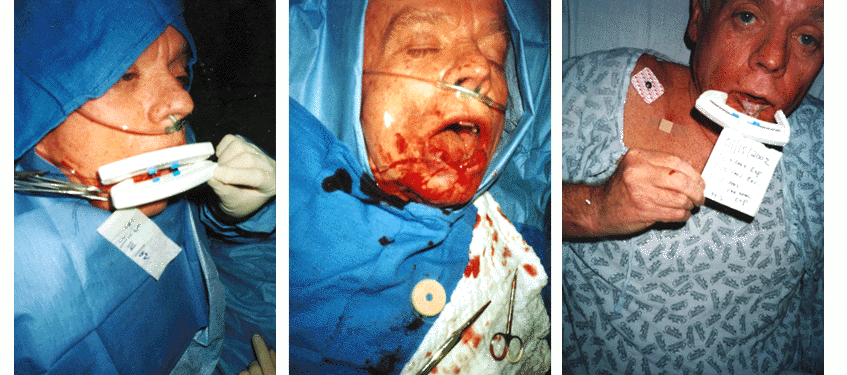
Photo 4 Photo 5 Photo 6
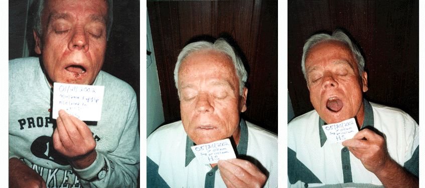
Photo 7 Photo 8 Photo 9 Photos Case 4
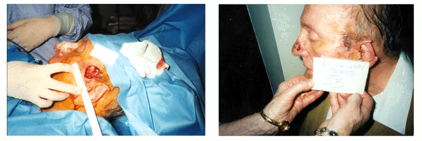
Photo 1 Photo 2
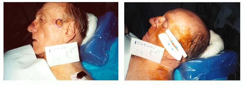
Photo 3 Photo 4
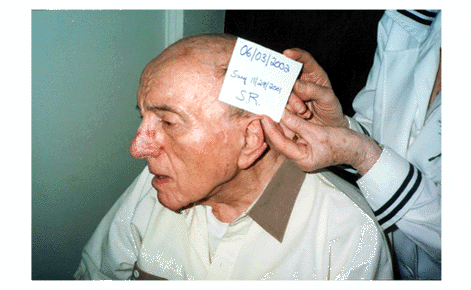
Photo 5
View Synopsis (.doc format, 365.0 kb)
See more of Posters
Back to Plastic Surgery 2003 Complete Scientific Program
Back to Plastic Surgery 2003 Meeting home
