Tuesday, October 28, 2003
3619
Anatomic Analysis of Surgical Approaches to the Deep Volar Forearm
PURPOSE: Forearm compartment syndrome threatens life and limb. The treatment is fasciotomy without delay, yet the standard surgical approaches do not address access to the deep compartments. If the deep space is not explored, contracture and/or necrosis may occur, with devastating results. Indications for evaluation of the deep space include electrical injuries, severe crush injuries, positional compression in comatose patients, and failed previous superficial fasciotomy. Deep fasciotomy often involves blunt, blind dissection, which places vascular pedicles and motor branches at risk for injury. Surgical disruption may contribute to ischemia and edema, adding to any injury already caused by the elevated tissue pressures. The goal of this study was to assess the ability of different surgical approaches to visualize the deep muscles without iatrogenic injury to the muscles, arteries, and nerves.
MATERIALS AND METHODS: Detailed anatomical analysis of 10 cadaver arms, two of which were injected with silicone and mildly plastinated. Findings were corroborated by live magnetic resonance angiography using a 3D fast spoiled-gradient echo technique that showed small vessels entering muscle. Three standard surgical approaches to the deep forearm were evaluated on each cadaver: radial (modified Henry and Spinner approach), central, and ulnar (modified McConnell approach). Iatrogenic injury to muscle, arteries, and nerves was evaluated on a scale of 1 – 3 (best – worst) for each category. Muscle injury was graded according to whether no dissection, sharp dissection, or division of the muscle was required for access to the deep space. Arterial injury was graded according to the number of segmental branches or dominant pedicles that required division. Nerve injury was graded similarly. The lowest possible score, corresponding to the safest approach, was 3. The highest possible score, corresponding to the most injurious approach, was 9. A Mann-Whitney Wilcoxon test for non-parametric data was used to analyze the data.
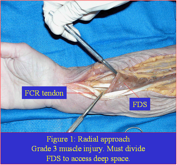
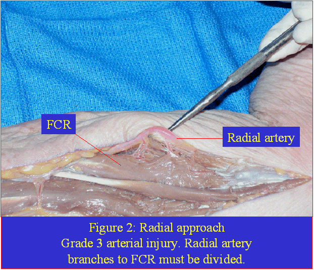
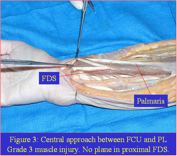
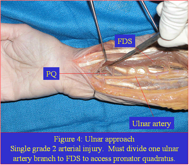
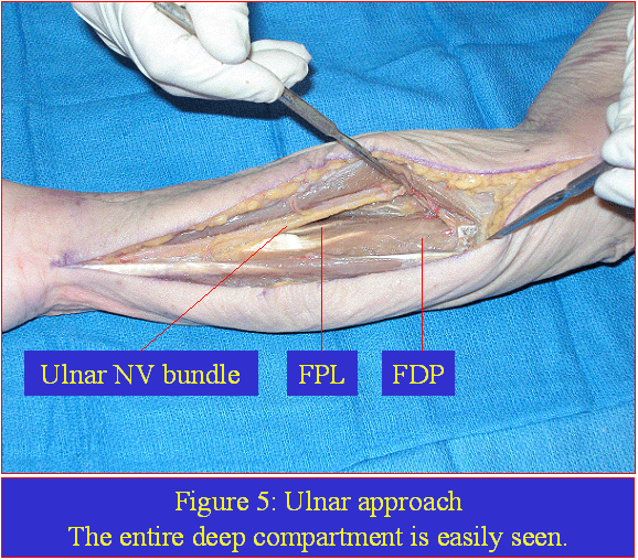
SUMMARY OF RESULTS: The radial approach scored an average of 6.1 (SD 0.32). Two significant findings were that the FDS III impeded access to the proximal deep space (Figure 1), and that several radial artery segmental branches to the FCR -- a Nahai-Mathes type IV muscle -- also impeded access (Figure 2). The central approach scored 5.8 (SD 0.63). There were two significant findings: the common musculotendinous portions of the FCR, FCU, and PL in the mid-forearm could not be easily separated (Figure 3), and if a portion of these muscles were opened surgically, the proximal musculotendinous portion of the FDS impeded access to the deep space. The ulnar approach scored 4.0 (SD 1.05). Only one ulnar segmental branch to the FDS required division (Figure 4). The entire deep space could easily be visualized (Figure 5). The ulnar approach caused significantly less iatrogenic injury than did radial and central approaches, with p values of 0.0001 and 0.0003, respectively. See examples below.
CONCLUSIONS: The ulnar approach to the deep compartment of the forearm was superior to other approaches. Other surgical approaches increase the risk of iatrogenic injury. Although none of the approaches is claimed to be clinically superior, the ulnar approach makes the most anatomical and logical sense.
View Synopsis (.doc format, 572.0 kb)
See more of Hand, Upper Extremity/Microsurgery
Back to Plastic Surgery 2003 Complete Scientific Program
Back to Plastic Surgery 2003 Meeting home
