
McCormick Place, Lakeside Center
Sunday, September 25, 2005
9:00 AM - 5:00 PM
McCormick Place, Lakeside Center
Monday, September 26, 2005
9:00 AM - 5:00 PM
McCormick Place, Lakeside Center
Tuesday, September 27, 2005
9:00 AM - 5:00 PM
McCormick Place, Lakeside Center
Wednesday, September 28, 2005
9:00 AM - 5:00 PM
8381
The Use and Mechanism of Action of High Intensity Focused Ultrasound for Adipose Tissue Removal and Non – Invasive Body Sculpting
Purpose: High Intensity Focused Ultrasound (HIFU) has been used extensively in the clinical setting to treat different types of tumors. However, there is no published literature on the use of HIFU for adipose tissue removal. The purpose of this study was to document the feasibility of use and mechanism of action of HIFU for adipose tissue removal and non – invasive body sculpting.
Materials and Methods: Initial transcutaneous HIFU therapy (Liposonix Prototype System, Liposonix, Inc., Bothell, WA.) was administered to the subcutaneous abdominal adipose tissue of swine. Once safety was established in the animal model, twenty four patients underwent HIFU treatment to their lower abdominal adipose tissue followed by subsequent abdominoplasty performed at different time points after HIFU therapy. The human clinical study was performed between July 15, 2003 and February 2, 2004 at Hospital Santa Monica in Mexico City after Ethics Committee and Ministry of Health Approval was obtained. Lesion fields from both the swine and human adipose tissue were examined for gross and histologic HIFU induced pathophysiological changes at various times after HIFU therapy, which ranged between several hours to eight weeks.
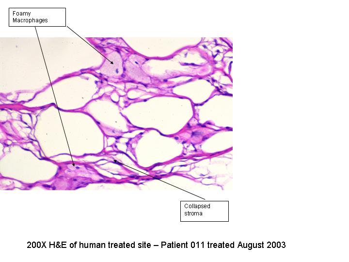
Results: Gross pathology revealed discrete lesion fields within the adipose tissue. The depth of each lesion was limited to the adipose tissue and did not extend into the dermis or the rectus muscle or fascia. The depth from skin to lesion field as well as the depth of the HIFU treatment site is determined by the transducer focal length. Peri-acute (hours) and acute (7 days) phase histology revealed a well demarcated zone of adipocyte disruption with minimal inflammatory response consisting predominantly of macrophages. Four week chronic histology revealed scavenger macrophages with abundant foamy cytoplasm within treatment zones. Eight week chronic histology demonstrated 75% resolution of the treated adipose tissue with collapse of the surrounding fibrovascular stroma. Lipid panels drawn from human patients throughout the trial did not reveal any new clinical changes from baseline. One week and six week full body necropsy studies on swine revealed no fatty liver changes or other systemic abnormalities after HIFU treatment.
Conclusions: The use and mechanism of action of HIFU therapy for fat removal has been proven in both pre-clinical and human clinical trials and provides a non- invasive method for body sculpting.
View Synopsis (.doc format, 106.0 kb)