
McCormick Place, Lakeside Center
Sunday, September 25, 2005
9:00 AM - 5:00 PM
McCormick Place, Lakeside Center
Monday, September 26, 2005
9:00 AM - 5:00 PM
McCormick Place, Lakeside Center
Tuesday, September 27, 2005
9:00 AM - 5:00 PM
McCormick Place, Lakeside Center
Wednesday, September 28, 2005
9:00 AM - 5:00 PM
8409
Finite Element Analysis of Fixation Plates for Mandibular Fracture Stabilization
Objective: To use finite element analysis (FEA) for evaluation of the biomechanics of plates used for the repair of mandibular fractures.
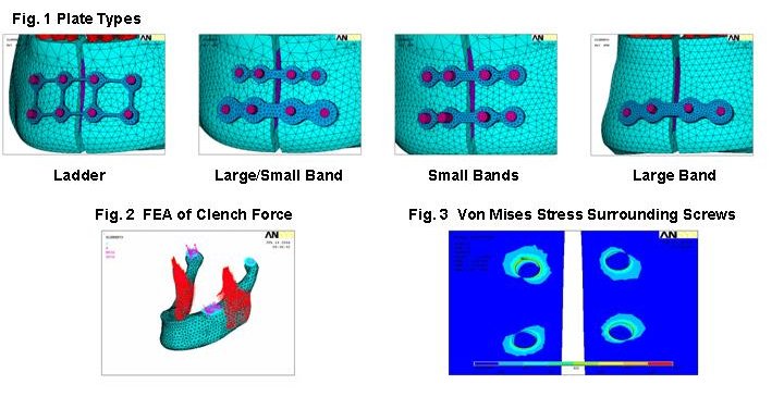
Methods: Computerized tomography (CT) data for the normal human mandible were obtained from a patient's craniofacial region imaged in the axial plane at 1.5 mm intervals. These data were then imported into Mimics 7.3 (Materialise, Glen Burnie, MD, USA) in image format. Masks for cortical bone, cancellous bone and dentin were designated separately with tools available in Mimics. The masks for each entity were approximated by IGES curves and imported into ANSYS 8.0 (Ansys Inc., Canonsburg, PA, USA). The volumes created for these entities were meshed using tetrahedral shaped solid elements. Each entity was modeled as elastic and isotropic. Data for ultimate tensile strength, Poisson's ratio and Young's modulus for each entity were taken from information on the material properties of the mandible available in the literature. An artificial gap was created in the symphysis of the mandible to mimic a fracture. Plates were created and shaped to the contour of the mandible overlying the region of the fracture using Solid Works 2001 Plus (Solid Works Corporation, Concord, MA, USA). The plates were then exported in IGES format into the pre-existing model of the mandible in ANSYS. Four plate configurations were compared for fitness of use (Fig. 1): ladder; large/small band; small bands; and large band. Unicortical and bicortical screws were simulated as solid cylinders. The force of the unilateral molar clench used for FEA was 150 N (Fig. 2).
Results: All bone-screw-plate constructs showed peak Von Mises stress surrounding the superior border of the screw proximal to the fracture on the side ipsilateral to the clench. Stresses did not exceed the ultimate stress for bone (120 MPa) in the region surrounding the screws on the anterior cortical surface. Stresses along the screw decayed towards the cancellous bone along the screw-bone interface. All configurations tested showed little or no stress in cancellous bone or lingual cortex. All configurations showed maximal tensile stress on the lower border of the mandible and maximal compression on the superior border.
Conclusion: On the basis of FEA, we conclude that all plate configurations tested are suitable to repair mandibular fractures. Unicortical screw fixation is adequate for fixation. Time required for contouring and fixation along with other ancillary features should be the principal criteria in selection of specific types of plates for mandibular fracture reduction.
View Synopsis (.doc format, 180.0 kb)