McCormick Place, Lakeside Center
Sunday, September 25, 2005
9:00 AM - 5:00 PM
McCormick Place, Lakeside Center
Monday, September 26, 2005
9:00 AM - 5:00 PM
McCormick Place, Lakeside Center
Tuesday, September 27, 2005
9:00 AM - 5:00 PM
McCormick Place, Lakeside Center
Wednesday, September 28, 2005
9:00 AM - 5:00 PM
8541
Perforator-based fasciocutaneous rotation flap
1. Purpose: To report our experience using perforator based fasciocutaneous flaps for buttock and lower leg reconstruction.
2. Introduction
Myocutaneous flaps have improved the management of buttock and the lower extremity soft tissue defects. However, there are several inherent disadvantages of muscle flaps such as functional deficits of the donor site and the bulkiness at the recipient site. To overcome these disadvantages, we used perforator-based fasciocutaneous rotation flaps for reconstruction of the buttock and lower extremity defects.
2. Material & methods
From March 2003 to February 2005 we operated on 14 patients using perforator-based fasciocutaneous rotation flaps. Ten flaps were based on perforators of the gluteus maximus muscle and 4 flaps nourished by perforators from the tibialis anterior and posterior system. The mean postoperative follow-up period was 1 year.
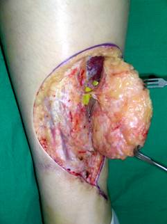
The technique involves localizing the flap perforators preoperatively with a Doppler. The flaps are elevated superficial to the fascia with preservation of one to three perforators. Rotation of the flaps is possible without dissection of the vessels deep to the muscle. The donor site is then closed primarily.
3. Results
All flaps completely survived and there were no perioperative complications. There was no functional disability of the donor area and the results were aesthetically pleasing.
4. Conclusion
Perforator-based fasciocutaneous rotation flaps for the reconstruction of buttock and lower extremity defects are an excellent alternative to musculocunteous flaps. The vascularity of the flaps is robust and dissection is technically easy. Perforator flaps do not require sacrifice of muscles, provide sufficient volume and are durable. Furthermore, these flaps result in less scar formation and allow more liberal dissection with safety. The donor defect can be closed primarily.
We conclude that perforator-based fasciocutaneous rotation flaps are very useful for reconstruction of the buttock and lower extremity.
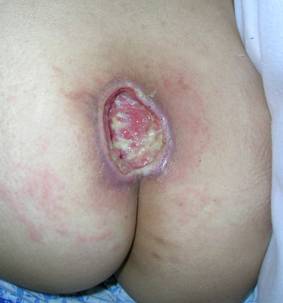
Fig 1. 30 years old female patient presented with chronic sacral sore (left).
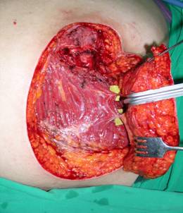
Fasciocutaneous perforator flap elevated on two musculocutaneous perforators (marked with vessel loops) (center).
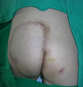
Postoperative 1 month view(right).
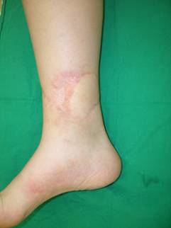
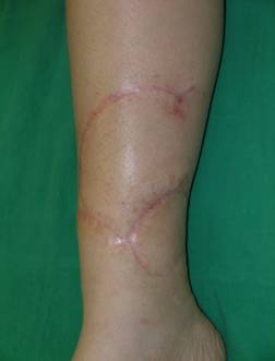
Fig 2. 13 years old male patient presented with an unstable soft tissue coverage of the distal tibia after partial necrosis of the previous island flap and secondary skin grafting (left).
Fasciocutaneous rotation flap supplied by one musculocutaneous perforator. (center).
1 month postoperatively (right).

