
McCormick Place, Lakeside Center
Sunday, September 25, 2005
9:00 AM - 5:00 PM
McCormick Place, Lakeside Center
Monday, September 26, 2005
9:00 AM - 5:00 PM
McCormick Place, Lakeside Center
Tuesday, September 27, 2005
9:00 AM - 5:00 PM
McCormick Place, Lakeside Center
Wednesday, September 28, 2005
9:00 AM - 5:00 PM
8606
Assessment of Distraction Regenerate Using Quantitative Bone Scintigraphy
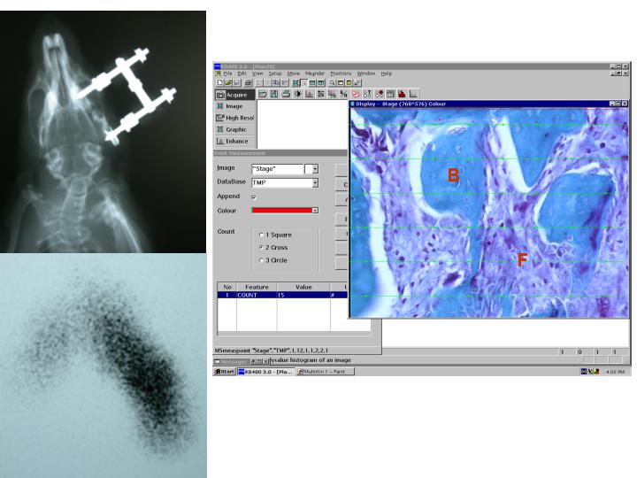
Distraction osteogenesis (DO) is a fascinating technique of endogenous tissue engineering that has become increasingly popular and revolutionized the treatment of numerous congenital malformations (1). Assessment of the newly formed bone after DO is the important step to test the effectiveness of therapy. Several clinical and experimental imaging techniques have been described for monitoring the newly formed bone during DO (2). Among these dual energy x-ray absorptiometry and quantitative computer tomography are more convenient method which can be used for large animal models (2). In small animal models assessment of distraction regenerate generally was performed with plain radiograph. However the potential of plain radiographs to provide information on callus formation and bone healing is limited and plain radiography has not been used successfully for quantification of bone healing (2). In this preliminary study we examined whether quantitative bone scintigraphy can predict the outcome of DO in small animal model. Materials And Methods: Recently we developed a rat mandibular distraction device that able to maintain its stability during the procedure and demonstrated that the device works properly and able to create ossified regenerate bones that filled the entire distraction gap, which can be used for various investigations about DO (3). This model was used in this preliminary study. Sixteen male Sprague–Dawley rats, weighing 250-300g, were used in this study. After a 3-days latency period, the right mandibles were distracted at a rate of 0,25 mm twice a day for 6 days. End of the activation period rats were divided into 2 groups. In group I (n=8) cephalograms were taken and quantitative bone scintigraphy and histomorphometric analysis (Figure-1) were performed at postoperative day 24 (second week of consolidation period). In group II (n=8) cephalograms were taken and the same analysis were performed at postoperative day 38 (fourth week of consolidation period). Quantitative Bone Scintigraphy: Before intravenous injection cephalogram was obtained and than 1 mCi of Tc-99m MDP was injected. Two hours after the intravenous injection of Tc-99m MDP, animals sacrificed and mandibles were dissected free of the surrounding tissues. Scintigraphic images which reflects the bone remodeling and mineralization were obtained using gamma camera equipped with pinhole collimator. By setting the region of interest on the distraction segment and contralateral normal area, the uptake ratio was determined. Following the bone scintigraphy mandible was prepared for histomorphometric analysis. Histomorphometric Analysis: The proportion of newly formed bone to fibrosis was analyzed on a microscope-based computer system which use a special image analysis software (Zeiss Vision KS 400 version 3.0 for Windows). Results: Uptake ratio was found over 3 for all osteotomy sites. Mean uptake ratios at 2nd and 4th weeks were found 5.95 and 4.08 respectively. The mean new bone formation at 2 weeks and 4 week were 57.3 ± 5.2 and 89.1± 4.7 respectively. Scintigraphic findings showed good correlation with radiographic and histomorphometric results. Conclusion: Quantitative bone scintigraphy is a promising method for the assessment of distraction osteogenesis and consolidation. It could offer objective qualitative and quantitative data for the noninvasive evaluation of bony regenerate.
References:
1. McCarthy J. G., Stelnicki E. J., Mehrara B. J., Longaker M. T. Distraction osteogenesis of the craniofacial skeleton.Plast. Reconstr. Surg. 107:1812-1827, 2001.
2. Swennen G. R. J., Eulzer C., Schutyser F., Hüttmann C., Schliephake H. Assessment of the distraction regenerate using three-dimensional quantitative computer tomography. Int. J. Oral Maxillofac. Sur. 34:64-73, 2005.
3. Eski,M., Nisanci,M., Cil,Y., Sengezer,M.,Ozcan, A. A Custom Made Distraction Device for Experimental Mandibular Distraction Osteogenesis. Accepted in J Craniofac Surg. .
Figure-1 : (Left, above) Cephalogram which was obtained at 2 weeks of consalidation period. (Left bottom) Scintigraphic images of the same mandible,(Right) Histological section,stained with Masson's trichrome, was assessed with image analysis software B indicates bone and F indicates fibrous tissue.
View Synopsis (.doc format, 381.0 kb)