
Sunday, September 25, 2005 - 10:05 AM
8715
Three-Point Fixation of Orbitozygomatic Fractures Without Skin Incisions: The Lateral Transconjunctival Approach to the Lateral Orbit
Introduction: Historically reduction and fixation of the lateral orbital wall and rim in orbitozygomatic fractures required a skin incision in the brow, lid, scalp, or canthal angle. In certain patients none of these approaches are ideal and can variously lead to unfavorable scars, distortion of delicate lateral canthal anatomy, or soft tissue ptosis. The medial transconjunctival approach to the medial orbital wall has recently gained popularity due to the excellent exposure provided and avoidance of cutaneous scars. We postulated that a lateral transconjunctival incision could similarly allow direct access to the lateral orbit while minimizing visible scarring.
Methods: A clinical and anatomic study was conducted. Twenty fresh cadaver orbits were dissected to elucidate the pertinent clinical anatomy of the lateral canthal region with respect to this new approach. The degree of exposure of the lateral orbital rim and wall through a 1.5cm lateral transconjunctival incision was assessed. Subsequent to completing the approach a detailed dissection was performed to evaluate whether visible damage had occurred to the levator palpebrae muscle, the lateral rectus muscle, Whitnall's ligament, the lacrimal gland, or tarsi.
In the clinical portion of the study we reviewed retrospectively our clinical experience with twelve orbitozygomatic fractures reduced and stabilized with the aid of the lateral transconjunctival incision. Outcome assessment included the use of post-operative computed tomography (axial, coronal and three-dimensional) to determine adequacy of bone reduction, and clinical assessment of lid position, conjunctival healing and soft tissue symmetry.
Results: The anatomic study demonstrated easy and complete exposure of the frontozygomatic suture and sphenozygomatic suture through a 1.5cm incision. Subsequent dissection showed the surrounding soft tissue structures to be undisturbed. Specifically the levator aponeurosis and its lateral horn, tarsi, lateral extension of Whitnall's ligament, lacrimal gland, and lateral rectus muscles were not visibly traumatized in any case.
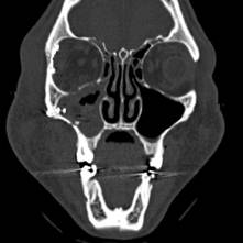
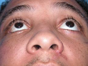
The clinical series of twelve fractured orbits was reviewed for complications and outcomes. Anatomic reduction was confirmed to be correct through post-operative computed tomography in all cases (Figure 1). There were no soft tissue complications (including ectropion, symblepharon, or ptosis of the malar fat pad) on clinical review at a minimum of six weeks post-operative (Figure 2).
Conclusion: Current surgical evolution in facial fracture care focuses on minimizing surgical morbidity while achieving the same standard of outcomes as traditional methods. The lateral transconjunctival approach to the lateral orbital rim and wall is anatomically sound and allows anterior three-point reduction and fixation of orbitozygomatic fractures without skin incisions when combined with intra-oral and/or inferior fornix incisions. This clinical study demonstrated the ability of this approach to allow anatomic reduction and fixation of orbitozygomatic fractures in twelve patients without soft tissue complications. Further investigation of this approach is deserved to define its role in the management of fractures involving the lateral orbit.ĀĀ
View Synopsis (.doc format, 55.0 kb)
See more of Cranio/Maxillofacial/Head & Neck
Back to 2005am Complete Scientific Program