
Monday, September 26, 2005 - 2:45 PM
9362
PSEF 2004 Research Fellowship - Lyndon Peer: Prevention Of Maxillary Collapse During Sutural Distraction Osteogenesis For Cleft Palate Closure
Introduction: Sutural distraction osteogenesis (SDO) of the longitudinal palatomaxillary suture has been suggested as a way to minimize the morbidity and scar burden of cleft palate closure. On review of the data from previous reports, however, the authors noted severe medial maxillary collapse associated with this technique. The authors hypothesize that 1) transverse distraction applied to the palatine bones achieves cleft palate closure preferentially by alveolar arch collapse, and not by intended SDO, and that 2) alveolar anchorage with a passive acrylic retainer could prevent arch collapse while achieving closure of palatal defects via SDO.
Methods: Palatomaxillary sutural distraction was carried out on 24, eight week-old growing beagle puppies with surgically-created palatal clefts. Three experimental groups were designated: controls (n=8), distracted animals without alveolar anchorage (distracted-only) (n=8), and distracted animals with alveolar anchorage (distracted-anchored) (n=8). Super-elastic NiTi closed-coil springs calibrated to 350 grams of force were used as distraction devices. The effects of distraction force on the sutures, palatal bone and maxillary complex were studied by intraoral measurements, craniometrics, gross pathology, qualitative and quantitative histomorphometry, 2D and 3D micro computed tomography (microCT), and Dual-energy Xray Absorptiometry (DXA).
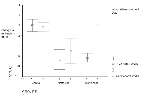
Results: Both intraoral measurements and craniometrics demonstrated severe, statistically significant decrease in anterior and posterior alveolar arch width in distracted-only animals, but not in distracted-anchored or control subjects (see graph - intraoral measurement data). Cleft gap closure was equivalent in both distracted groups. Histologic analysis revealed increased suture width and tortuosity at 2 weeks post-distraction in distracted-anchored which reverted to control group suture morphology by 12 weeks post-distraction. Quantitative histomorphometry and microCT both demonstrated a statistically significant increase in percentage bone composition around the distracted palatomaxillary suture in distracted-anchored animals versus controls, while DXA measured no detectable changes in bone mineral density (BMD) between the distracted and control specimens.
Conclusions: This study has shown that simple distraction applied to the palatine bones causes significant preferential arch collapse over sutural bone deposition. By utilizing an acrylic retainer for alveolar anchorage we were able to prevent arch collapse in this model and demonstrate a quantitative increase in bone deposition over controls with statistical significance. Furthermore, unaltered BMD and post-distracted suture morphology suggests that there is no structural compromise that may impact future growth or stability. Thus, this technique, with the described modification, could theoretically decrease the scar-related complications of cleft palate closure caused by mucoperiosteal undermining techniques currently employed in human infants.
See more of Technology and Research
Back to 2005am Complete Scientific Program