McCormick Place, Lakeside Center
Sunday, September 25, 2005
9:00 AM - 5:00 PM
McCormick Place, Lakeside Center
Monday, September 26, 2005
9:00 AM - 5:00 PM
McCormick Place, Lakeside Center
Tuesday, September 27, 2005
9:00 AM - 5:00 PM
McCormick Place, Lakeside Center
Wednesday, September 28, 2005
9:00 AM - 5:00 PM
8127
Cost-Benefit Analysis for Pathologic Examination of Reduction Mammoplasty Specimens
Background: Reduction mammoplasty has a number of indications, including relieving back pain, improving the cosmetic appearance of the breast, and providing symmetry after excision of a contralateral malignancy. Reduction mammoplasty is not indicated for resection of malignant or premalignant lesions of the breast. Despite this, the excised breast tissue is routinely sent for pathologic examination. Titley et. al. in 1996 reported a case series of 157 women undergoing reduction mammoplasties. Pathologic examination of all specimens failed to identify any malignant or premalignant changes. This raises into question the value and cost benefit of submitting breast reduction specimens to pathology. Our study seeks to determine the incidence of premalignant changes in the pathology of women undergoing reduction mammoplasty at our institution, as well as to determine the cost-effectiveness of pathologic examination of specimens from reduction mammoplasty. Specifically, the study examines whether it would be more cost-effective to only submit the specimens from women age 40 or above, as that is the age at which routine screening mammography is recommended.
Methods: A retrospective chart review was performed on 300 women who underwent bilateral reduction mammoplasty at a tertiary care center between 1991 and 1999. Any patient with a previous history of breast cancer was excluded. The inferior pedicle technique was used for all patients, with a total of four surgeons performing the operations. The specimens were sent to pathology in formalin, and were then cut in 1 cm intervals for gross inspection. If no gross abnormalities were seen, three sections from each breast were sent for histologic examination. If gross abnormalities were present, then more cuts from the specific area were obtained.
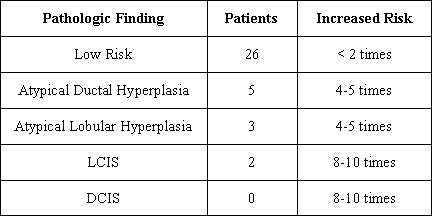
Results: In this study, 36 of the 300 patients (12%) had abnormal pathology reports which indicated either a premalignant lesion, or a lesion which puts the patient at increased risk of developing breast cancer. Seventy-two percent (26/36) were low-risk lesions. These included apocrine changes, duct ectasia, moderate or florid hyperplasia, sclerosing adenosis, or papilloma. Three percent of all patients in the study (10/300) had lesions which were moderate or high risk, and would put them at significantly higher risk than the general population for developing breast cancer. The average age of patients with benign pathology was 32.6 years old (range 14 to 67), and the average age of patients with abnormal pathology was 42.5 years old (range 16 to 73). The cost of pathologic examination of one breast specimen at our facility is $190.86 ($381.72 for a bilateral reduction mammoplasty), resulting in a total cost of $229,032 for all 300 patients. The cost to identify a patient with a moderate to high risk lesion was $22,903. If pathologic examination was restricted to women 40 years or older (86 patients), the total cost would have been reduced to $32,828, with a total savings of $196,204. In this study of 300 patients, 10 had moderate to high risk pathology. Of these 10 patients, 2 were less than 40 years old. These 2 patients showed LCIS and atypical ductal hyperplasia on their pathology reports.
Conclusions: Limiting pathologic examination of reduction mammoplasty specimens to women older than 40 years would fail to identify 20% of moderate to high risk pathology. Despite the potential cost savings, this is not an acceptable risk.

View Synopsis (.doc format, 36.0 kb)
