McCormick Place, Lakeside Center
Sunday, September 25, 2005
9:00 AM - 5:00 PM
McCormick Place, Lakeside Center
Monday, September 26, 2005
9:00 AM - 5:00 PM
McCormick Place, Lakeside Center
Tuesday, September 27, 2005
9:00 AM - 5:00 PM
McCormick Place, Lakeside Center
Wednesday, September 28, 2005
9:00 AM - 5:00 PM
8642
Dermal Wound Closure Method Alters Scar Structure
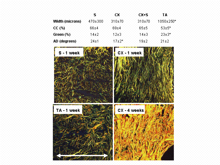
Background: Collagen plays a vital structural role in wound healing, with fiber content, thickness and orientation all influencing scar integrity. Our hypothesis was that the method of wound closure could alter these structural parameters. We tested this concept using 4 closure techniques. Methods: A 29 mm full-thickness incision was made on the back of 64 rats, randomized to closure with sutures (S), a coaptive film device (Clozex, CX: exerts tension perpendicular to the wound via adhesive pads and filaments), CX + deep absorbable sutures (CX+S), or tissue adhesive (TA; high-viscosity Dermabond). Histologic sections stained with picrosirius red were viewed with polarized light at 1 and 4 weeks (the sutures and closure devices were removed at 2 weeks). We assessed scar width, collagen content (CC), and fiber thickness (collagen color changes from green to yellow to orange as fibers mature and thickness increases). In addition, we measured the two-dimensional orientation of 50 collagen fibers in each scar and in the intact dermis, distant from the incision, in 4 samples and calculated the angular deviation (AD) of each distribution obtained (the smaller the AD, the more aligned the collagen fibers). Results: At 1 week (Table), TA-treated scars were wider (*P<0.05, ANOVA), contained less collagen (P<0.05) and a greater proportion of immature green fibers (P<0.05). At 4 weeks, scar width decreased (170-225 microns) and all groups displayed increased collagen content (94-95%) and fiber thickness: few green fibers remained (1%) and >60% of the fibers were mature (orange) (P=NS between groups). Collagen fiber orientation differed among closure methods; i.e., increased fiber alignment was seen with CX at 1 week (smaller AD; P<0.05). At 4 weeks, the fibers were less aligned and the average AD had increased (24.5 degrees); however, the organization was not the same as in the normal dermis, which had a near random alignment (AD = 35.0 degrees). The figure shows representative examples; in all cases, the incision runs horizontally as marked by the arrow – generally, fibers were aligned perpendicular to the incision. Conclusions: The method of dermal wound closure altered healing. Poor early closure with tissue adhesive delayed healing; the wider scars contained less collagen and a greater proportion of immature, thin, green fibers. In contrast, the film device provided more uniform wound closure and increased fiber alignment. Nevertheless, at 4 weeks, there was no difference between groups. Thus, the early advantage provided by film device did not compromise late healing.

View Synopsis (.doc format, 42.0 kb)
