
Sunday, October 8, 2006
10569
The Precision of High-Intensity Focused Ultrasound (HIFU) for Non-Invasive Body Sculpting: In Situ Measurement of the HIFU Treatment Zone within Adipose Tissue
Purpose: The therapeutic effects of HIFU are dependent on energy deposition in the target tissue, which produces cell death secondary to thermal coagulation of the tissues. Thermal effects from HIFU are the direct result of temperature increases from the deposition of ultrasonic energy along with heat generated within the cells and the interstitium from increased pressure within the targeted tissue. There is a steep temperature gradient between the HIFU treated tissue and the adjacent tissue that is not within the HIFU treatment beam. The purpose of this study was to perform in situ thermocouple measurements of HIFU treated adipose tissue of swine to evaluate the precision of the thermal effects, and tissue changes produced. The intervening and adjacent structures were studied for presence of unintended damage outside the treatment zone.
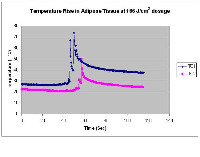
Materials and Methods: K-type thermocouples were positioned in the Yorkshire swine model at various anatomical locations including: epidermis, dermis, subcutaneous adipose tissue at depths of 10 and 15 mm, and within the intra-abdominal cavity. The skin directly above the thermocouples was marked. Placement depth of the thermocouples was verified by ultrasound. Transcutaneous HIFU at dosages between 166 J/cm2 and 372 J/cm2 (Liposonix Prototype System, Liposonix, Inc., Bothell, WA.) was administered at a focus of 15mm beneath the skin at the site where the thermocouples were placed in the adipose tissue. Temperature data from the thermocouples was collected every 500 msec during the duration of the treatment and for several seconds after the completion of therapy using an Agilent 34970A Data Acquisition/Switch Unit and a laptop PC.
Results: The temperature measured in the abdominal cavity did not deviate from 36.8°C while treating the site at dosages between 166 J/cm2 and 372 J/cm2. There was an acute temperature rise to over 70°C at the focal beam within less than 1 second (sec). There was no temperature elevation in the epidermis or dermis or superficial adipose tissue layers as recorded by the superficial thermocouples. Once the focal point of the HIFU beam has passed by the thermocouple in the tissue, there is a gradual temperature decay of greater than 30 °C. Post- treatment pathology demonstrates no damage to the intervening or adjacent structures outside of the treatment zone.
Conclusions: The ability to concentrate the focused ultrasound energy within the targeted tissue prevents thermal scattering to tissues outside of the focal zone. The temperature at the treatment zone and the volume of tissue treated can be precisely controlled depending on the dosage used. In addition, there is no mechanical damage from cannula insertion as there would be from lipoplasty, thus limiting tissue damage to the target area only. The precision of HIFU provides a practical and controlled method for performing non-invasive body sculpting.
View Synopsis (.doc format, 32.0 kb)