
Sunday, October 8, 2006
10723
Reconstruction of Failed VAC Closure with External Skin Expansion
Reconstruction of Failed VAC Closure by External Skin Expansion.
Purpose.
To demonstrate tension free abdominal wall reconstruction with external skin expansion after failed VAC treatment. All wounds must be cleaned, cut, closed or covered (Schessels 4Cs). The wound must be free of piss, puss. poop and particles (Schessels 4Ps)
Method
At 1st surgery wound margins are approximated by 20 nylon sutures on a PSLX needle placed 2 cm apart and 2 cm from wound margins. One suture is tied, the ends left 7 cm long, adjacent suture is left untied. Untied suture will be tied at 2nd application of expanders. Expanders are applied and secured to patient by long ends of suture. This process is repeated until an abundant amount of tension free tissue is accumulated. At 2nd surgery wound margins are freshened, closed by suture and a catheter is placed for subcutaneous irrigation.
Case 1
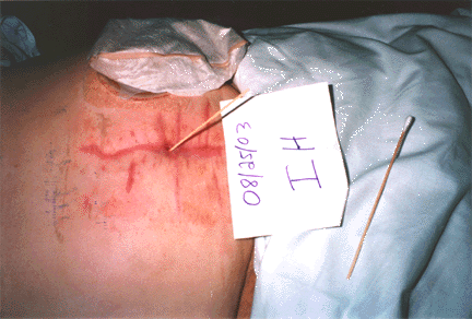
Photo 1 - A 73 year old patient had an exploratory laparotomy for a perforated diverticulum on 3/13/03. Two attempts to close wound failed. Wound closed after 3 months of VAC treatment leaving a deep draining infected fistula.
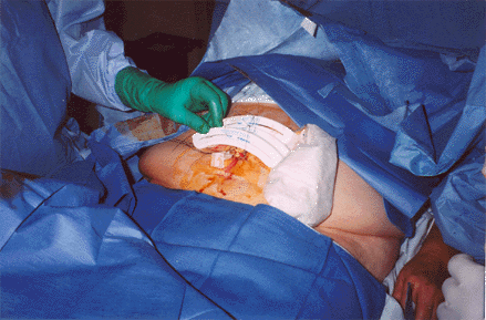
Photo 2 – Patient admitted on 8/25/06 - 1st Surgery application of external expanders
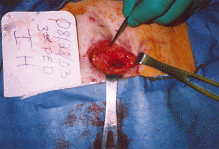
Photo 3 –After 3 days external expansion - 2nd Surgery Excision of deep fistula and wound is closed by suture.
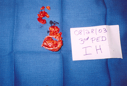
Photo 4 – Excised deep fistula
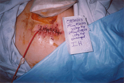
Photo 5 – Irrigation catheter placed for deep subcutaneous irrigation for 7 days.
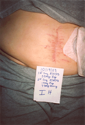
Photo 6 –Appearance 6 weeks post closure
Case 2
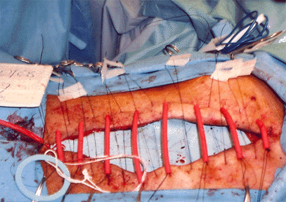
Photo 1 – 82 year old patient admitted with an acute abdomen. Surgery was performed on his gall bladder. Post-op there was severe abdominal pain, distension and an elevated temperature of 104+. On the 2nd post-op day an exploratory surgery revealed a ruptured appendix, abdomen was left open, wound placed on a VAC for 2 ˝ months. Wound failed to heal, wound measured 14.3 by 12.9 cm.
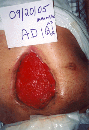
Photo 2 – At 1st surgery a protective fish barrier is placed and wound margins are approximated by suture.
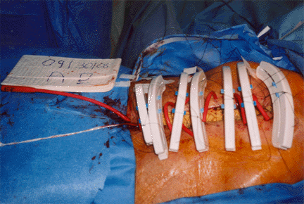
Photo 3 – Expanders are applied.
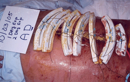
Photo 4 – Appearance 3 days expansion.
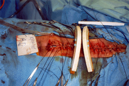
Photo 5 – At 2nd surgery, wound edges are freshened, wound is closed by suture, a catheter is placed subcutaneously and expanders are applied for additional day.
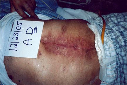
Photo 6 – Appearance at 10 weeks.
Total cases – 4
Conclusion
VAC treatment is a slow method to clean and close wounds. When VAC therapy fails, external expansion is indicated. Wounds are best closed with tension free, well vascularized thick local skin and subcutaneous tissue. This method can also be used for primary closure of contaminated wounds following extensive surgical debridement, irrigation and tissue expansion. Tissue expansion causes an increase in angiogenesis. Most procedures are carried out under local or MAC anesthesia.
View Synopsis (.doc format, 557.0 kb)