
Sunday, October 8, 2006
10836
A Study on Blood Flow in Pedicled Jejunum after Supercharge Using a Dog Model -Which is More Important for Survival of Distal (Oral) End of Pedicle Jejunum, Artery or Vein? -
Background: Pedicled jejunum and colonic interposition are very useful for thoracic esophageal reconstruction especially in case where a gastric tube is unavailable. However, partial graft necrosis of the oral end of transferred intestine can occur due to poor circulation. Microvascular augmentation (supercharge) of the jejunum to improve circulation has been performed with good clinical results. However, there are very few reports about the tissue blood flow of the pedicled jejunum and which is more important for survival of distal (oral) end of pedicled jejunum, artery or vein.
Material and method: To verify this, the authors performed an experimental animal study using dog jejunum and measured the tissue blood flow using laser flowmetry. Adult female northern beagle dogs, weighing 8-10kg, were used in this study. Under genernal anesthesia, first, second and third jejunal vessels were identified after laparotomy.
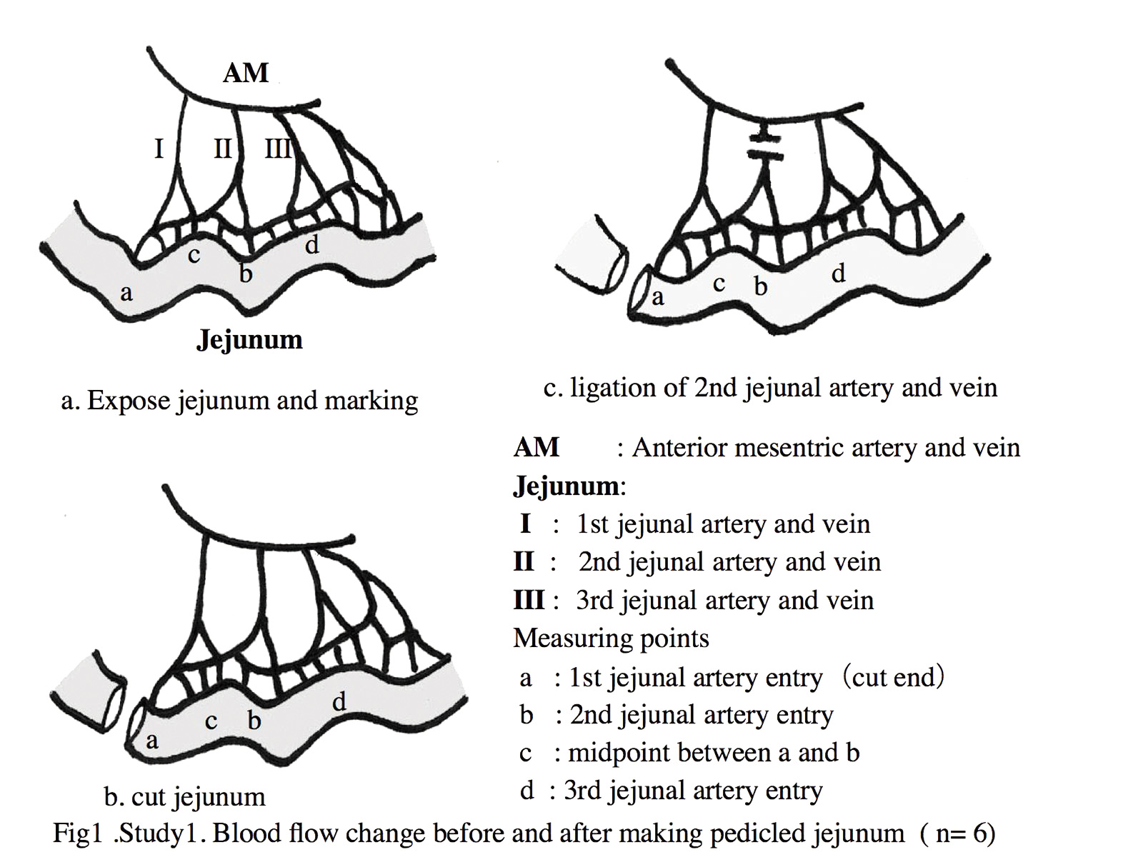
Study 1: Point a, point b and point c were marked using 5-0 nylon stitches. (Fig.1a). Point a is opposite and more oral side the influx point of first jejunal vessels. Jejunum was intended to be cut at this point. Point b is opposite the influx point of second jejunal vessels and point c is mid point of point a and point b. Point d, opposite influx point of third jejunal vessels, is also marked. The tissue blood flow was measured at point a, point b and point c Then jejunum was cut at more oral side at point a and tissue blood flow was measured at point a, b and c (Fig. 1b). After this procedure, second jejunul vessels were ligated (Fig. 1c) and tissue blood flow were measured at these three points. In six dogs, this study were performed. Tissue blood flow change at every point and difference between point a ,b and c were evaluated by Wilcoxon signed lank test.
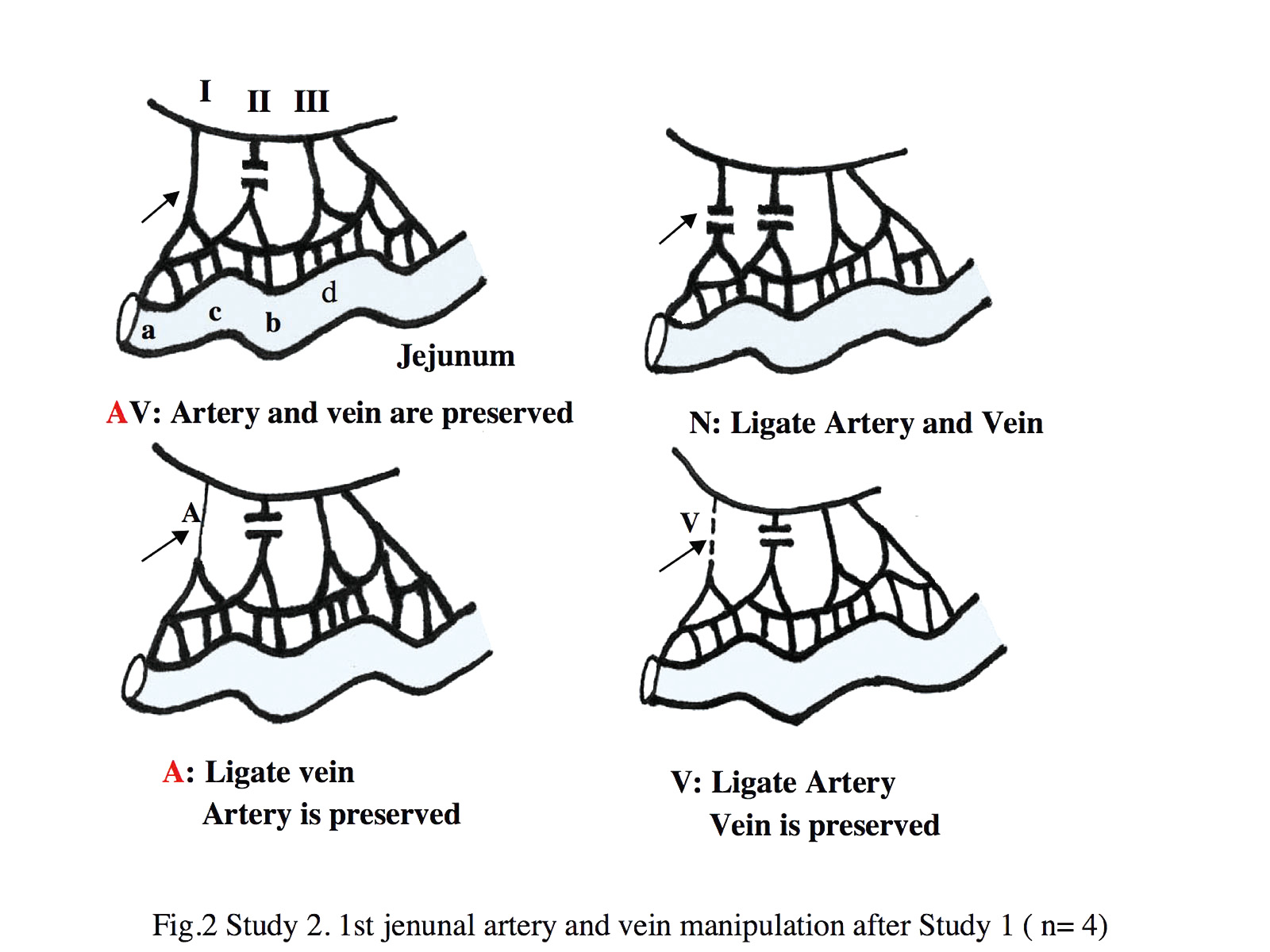
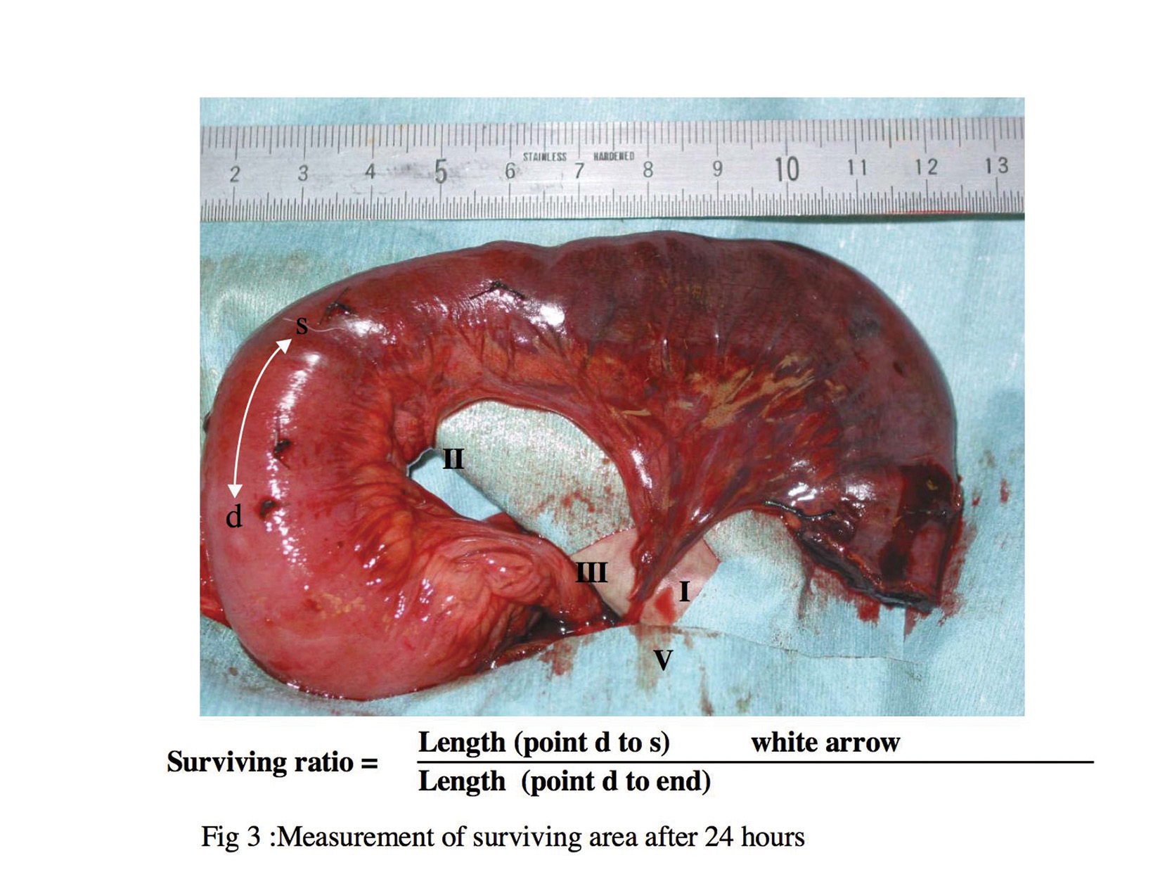
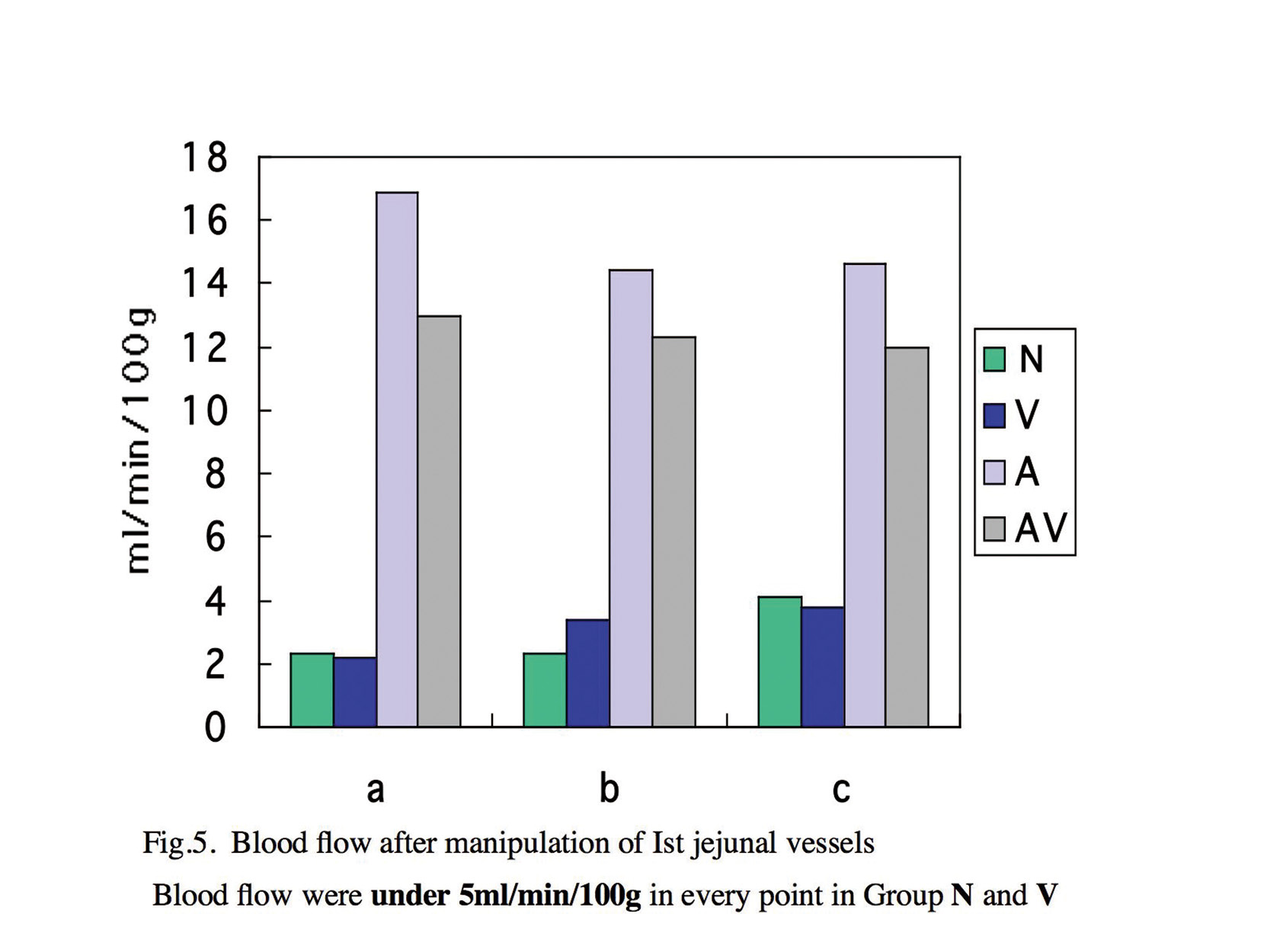
Study 2:After measurement, first jejunal vessels was manipulated to determine which vessel is more important, artery or vein, for tissue blood flow in distal (oral) side of pedicle jejunum. We divided animals into four groups to compare blood flow and tissue survival area under different supercharge conditions (Fig 2). Group AV: First jejunal artery and vein were only divided. Both vessels were preserved. Group A: First jejunal artery was preserved but first jejunal vein were ligated. Group V: First jejunal vein was preserved but first jejunal artery was ligated. Group N: Both first jejunal artery and vein were ligated. Every group had one dog. Tissue blood flow were measured before and after manipulation at point a, b and c. After measurement, both edge of jejunum were sutured as stoma and wound were closed. Animals were feed intravenously, then were re-anesthetized after 24hours to examine surviving area of pedicled jejeunum in every group. Edge of survival area was marked as point s. Surviving area was measured in every group and survival ratio was calculated. Surviving ratio = survival distance (point d to point s) / distance from point d to edge of pedicle jejunum (Fig.3).
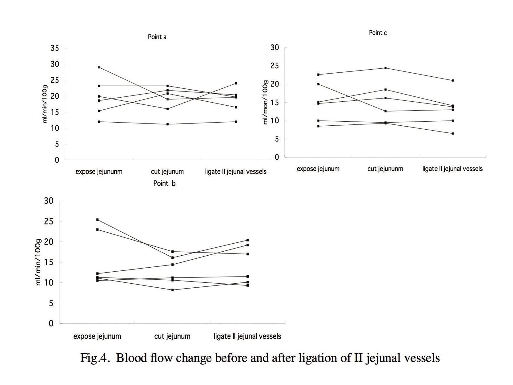
Results: In study 1, there were no signignifinant difference in blood flow between point a,b and c.
There were no significant difference in blood flow accorcing to procedure in study 1(Fig.4).
It indicates that ligation of the dominant vessel doesn?ft decrease the perfusion significantly as the jejunum will be nourished continuously by the adjacent jejunal vessels.
In study 2, blood flow at point a, b and c decreased significantly in Group N and Group V after manipulation of first jejunal vessels(Fig.5). However, in Group A and Group AV, blood flow did not decrease. Survival ratio was 100% in Group A and Group AV 24hours after manipilation of first jejunal vessels, whereas 20% in Group V and 21% in Group N .
Our study showed that arterial anastomosis is more important than the venous anastomosis for the survival of distal (oral) end of the supercharged pedicled jejunum.
View Synopsis (.doc format, 492.0 kb)