
Sunday, October 8, 2006
10896
Novel Approach to Close Treatment of the Prominent Ear Without Skin Resection (Endotoplasty)
The aim of this study is to demonstrate an alternative closed approach to treat prominent ear, without skin resection, performed through a single 5-8mm incision located on the external border of the helix at the exit of the posterior crura. The strategy is delineated by anterior and posterior global undermining of the auricular skin over the anti-helix and the concha; controlled anterior anti-helix rasping with stabilization of the antihelical row using trans-cutaneous stitches, and resection of the hypertrophic concha according the “conchal show” principles.1 Considering the minimum access, preservation of the auricular structure and the closed modelation of the auricular cartilage, the term endotoplasty is proposed for this procedure.
PATIENTS AND METHODS
The surgery was performed on 342 patients ( 214 female and 128 males, mean age 22.7 years), and included 328 bilateral and 14 unilateral cases(n=670 ears), over a period of five years. The data was classified according to the gradation proposed by the Egloff et all.2 Based on this classification, 178 patients belonged to the type I(altered anti-helix associated with conchal hypertrophy), 6 patients to the group II( hypertrophied concha and normal anti-helix), 132 to the group III(altered anti-helix, normal concha), and another 26 to the group IV (association with one of the former variation with a lateral lobe).
RESULTS
A balanced external ear configuration was achieved in majority of the cases. A smooth antihelix surface was consistently achieved with few complications. The most frequent complication was sinus formation around the knots of the trans-cutaneous stitches and occurred in 12 patients(3,5%). This inconvenience reduced dramatically, when the stitches were done with either unabsorbable 5-0 clear nylon (Ultralon®- Biosut-MG) or absorbable 5-0 PDS(Sommerville-Eticon®). Moderate asymmetry were present in 5 patients (1,4%). It was correct utilizing the same strategy. Hematoma occurred in 3 patients(0,8%). No infection or pathologic scar were registred. The mean follow-up period ranged from six months to five years.(Fig. 3,4)
In order to measure the patient satisfaction, the Caouet-Laberge et all.3 questionnaire was applied. 98% of the patients scored to be very satisfied or satisfied.
DISCUSSION
In this investigation, a closed method to correct the prominent ear is evaluated. The entire closed procedure can be performed through a single 5-8mm incision located on the external border of the helix and the exit of the posterior crus. The strategy starts with mandatory tumescent infiltration, to facilitate the undermining over the anti-helix and the concha, both in front and behind the ear. The cartilaginous mold stays free to be sculptured. The global schrinkage of the skin envelope contributes to the medianization of the auricle, making skin resection unnecessary. The instrumentation is composed of a skin descolator, one direction thin rasp, (similar to the one utilized to smooth the nasal dorsum) and one full curved Fomon pair of scissors . Eletrocautery is not utilized. The surgeon can accomplish the whole treatment without auxiliar.
The hypertrophic concha present in the types I, II and IV had to be corrected in more than 60% of the auricles. To address this problem, we embodied the concept of “conchal show” advocated by Vermellian and cols. They analyzed 100 patients with clinically normal ear, recording the ideal conchal width, which varied from 8 to 11mm. We considered conchal width of 10mm as the limit. The excess is resected in order to promote medianization of the auricular
CONCLUSION
The closed approach, performed through a single small incision on the external border of the helix, at the union of the exit of the posterior crus, can correct effectively prominent ear types I, II and III of the Egloff grading. This strategy requires tumescent infiltration with anterior and posterior undermining of the auricula; controlled anterior scratching of the anti-helix with its stabilization using trans-cutaneous stitches and correction of the hypertrophic concha according the “conchal show” principles.
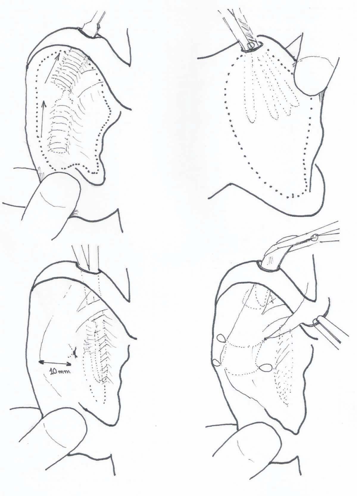
Figura1
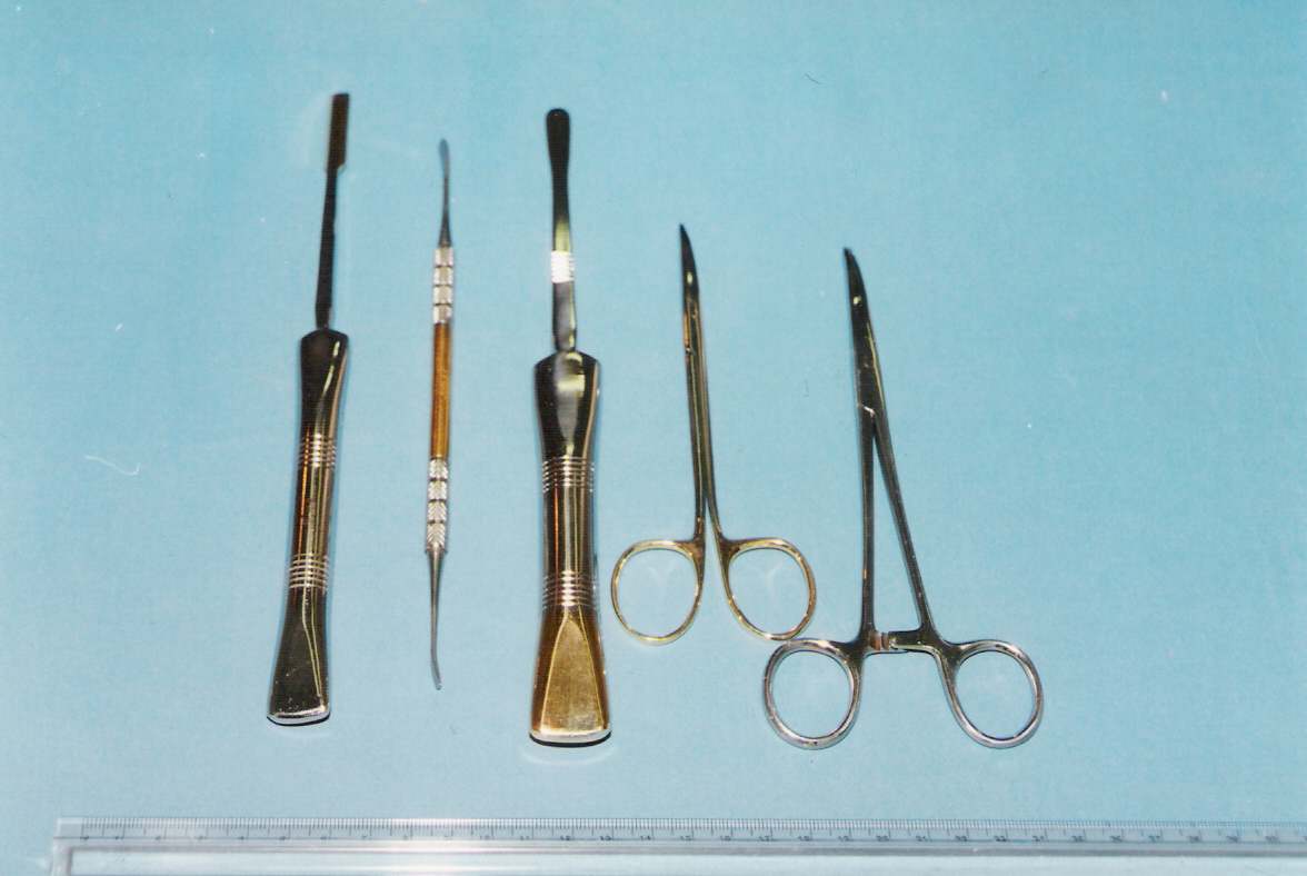
Figura 2
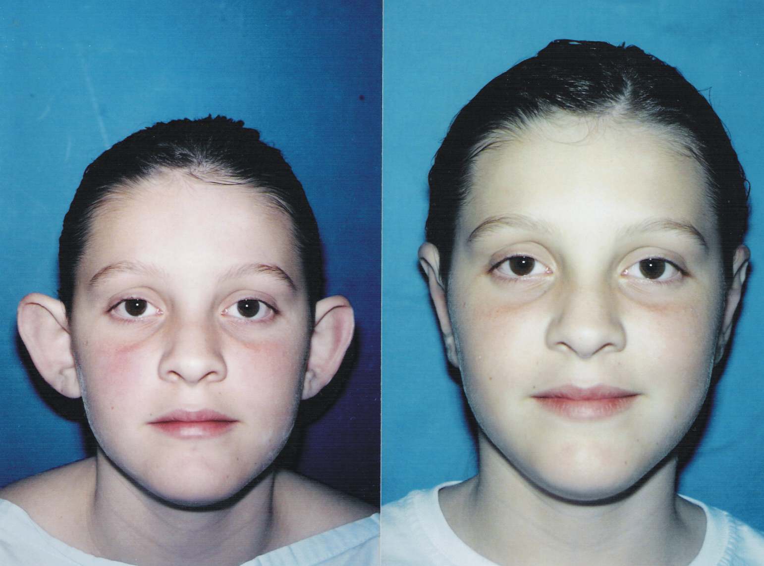
Figura 3

Figura 3 – Close
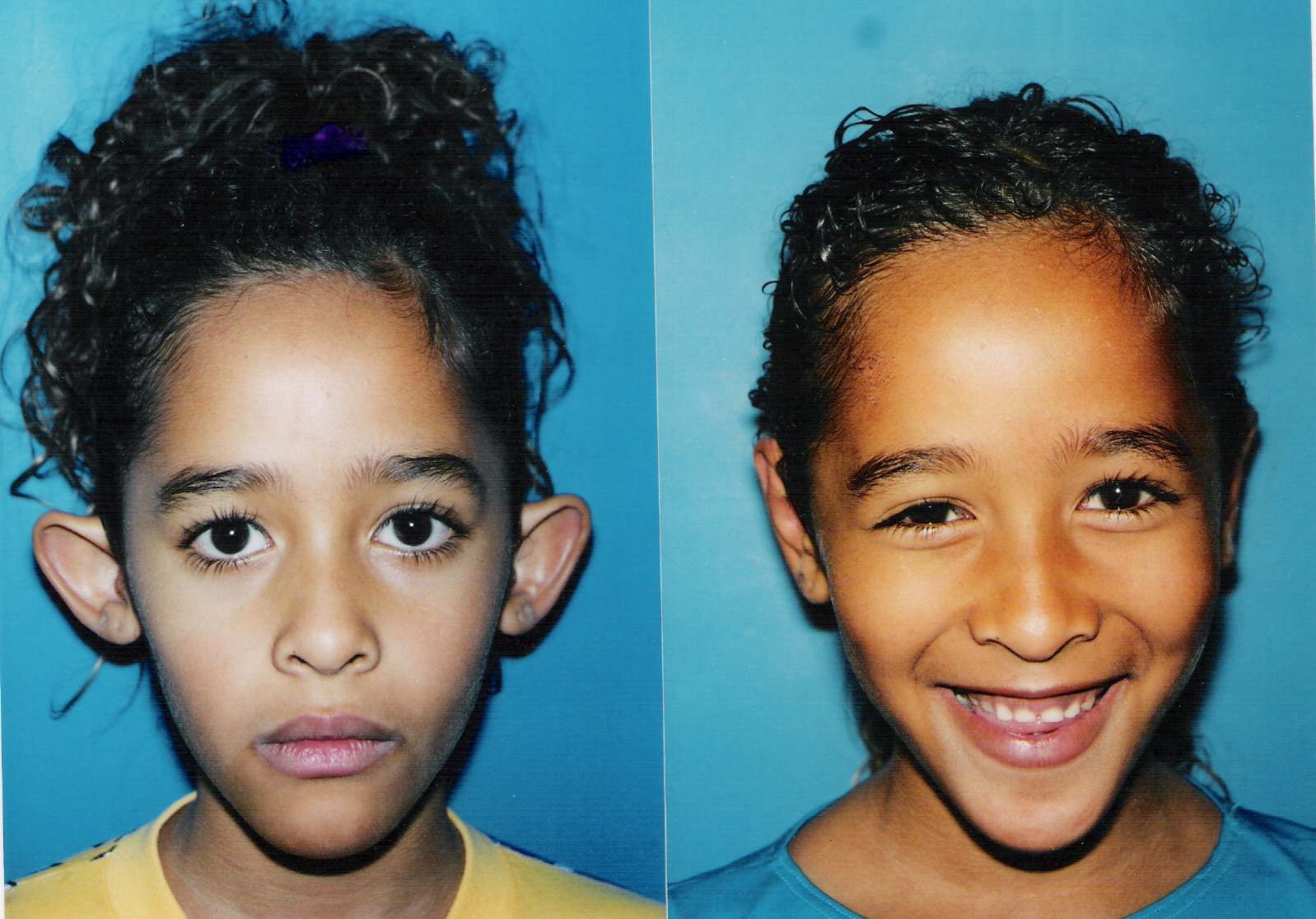
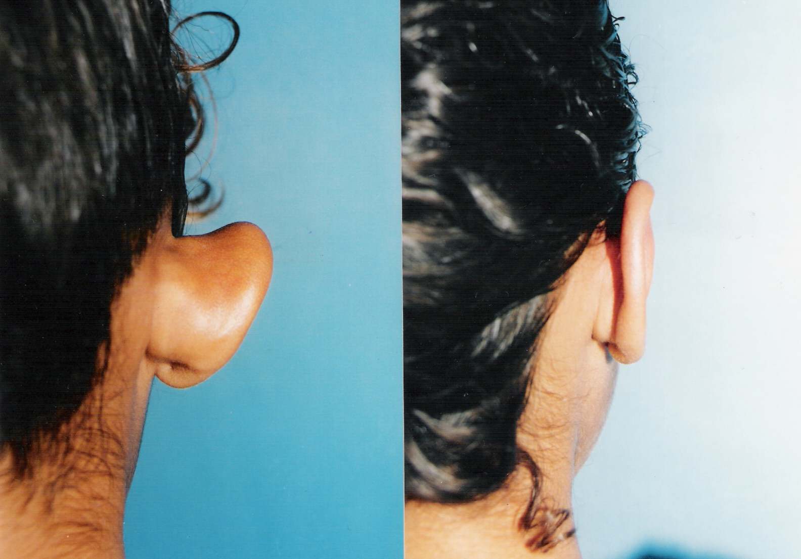
Figura 4

Fig 5
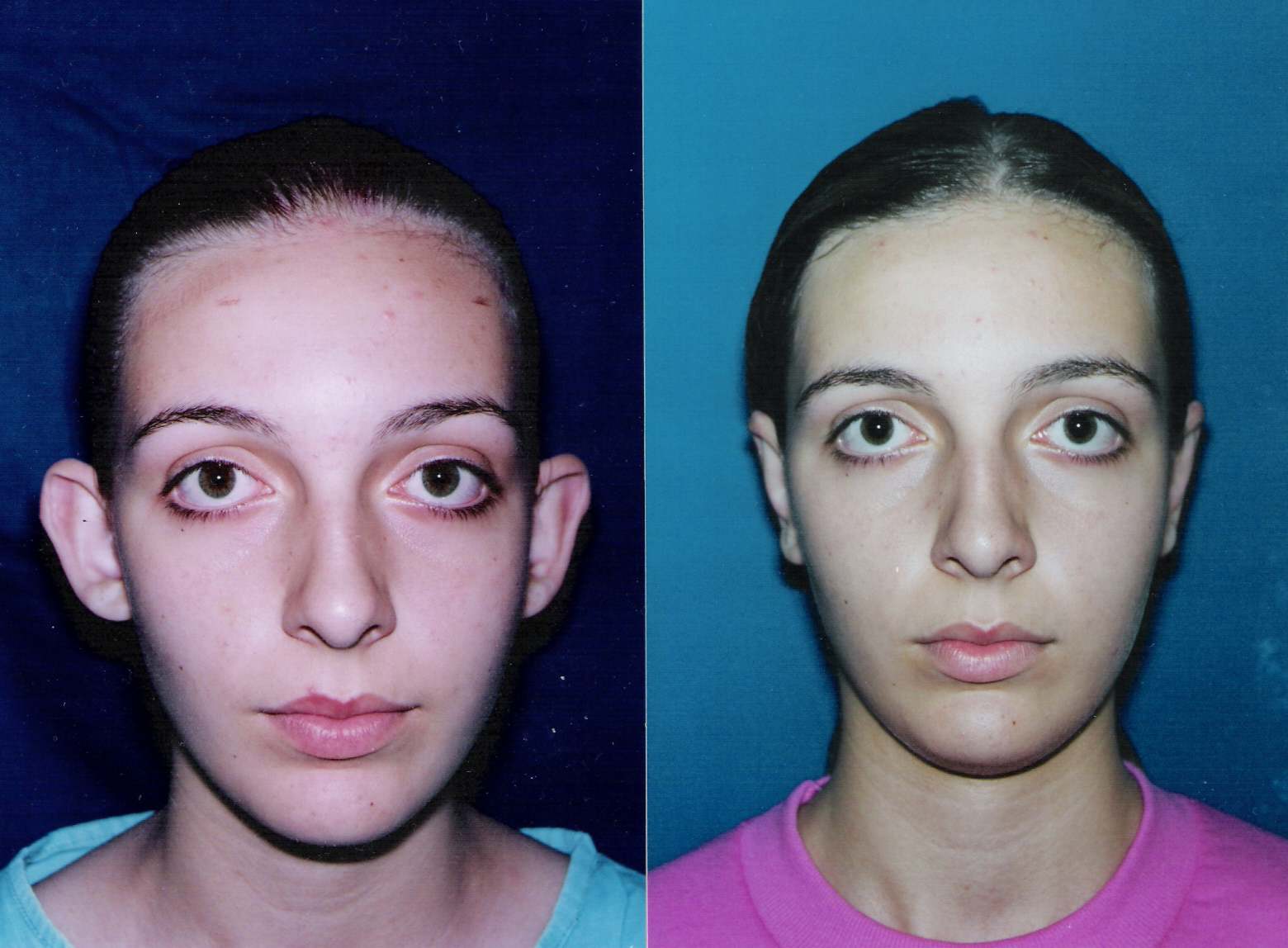
Fig. 6
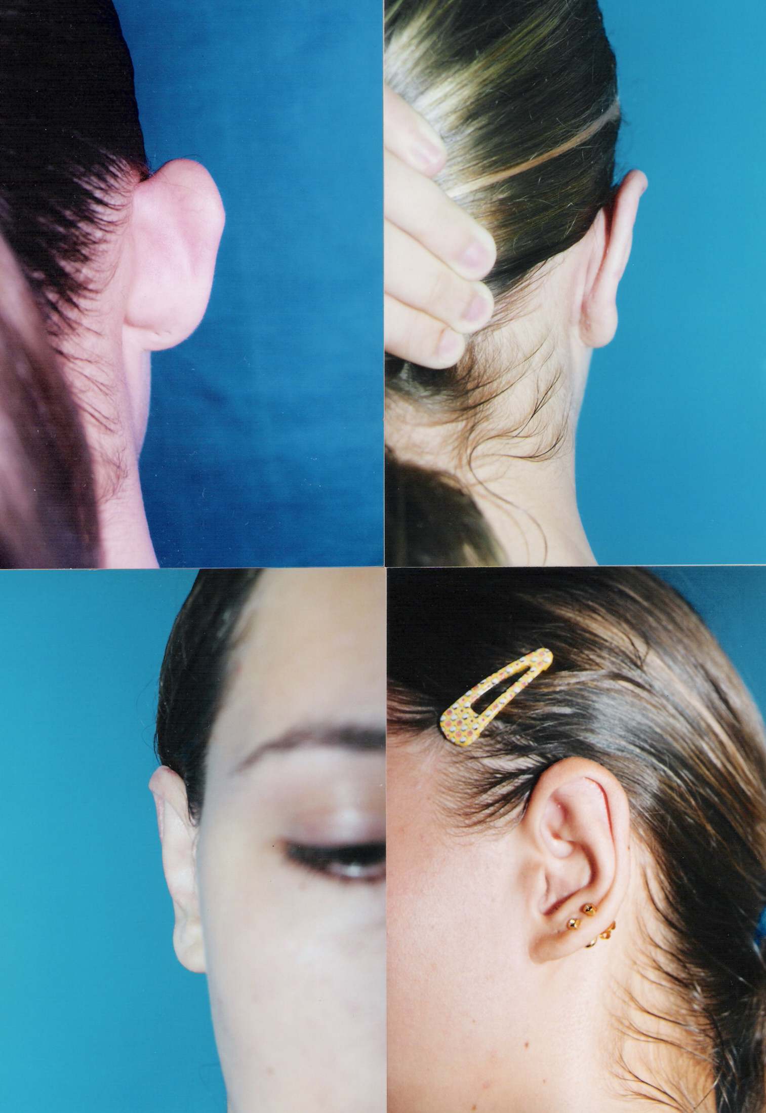
Fig. 6 - close
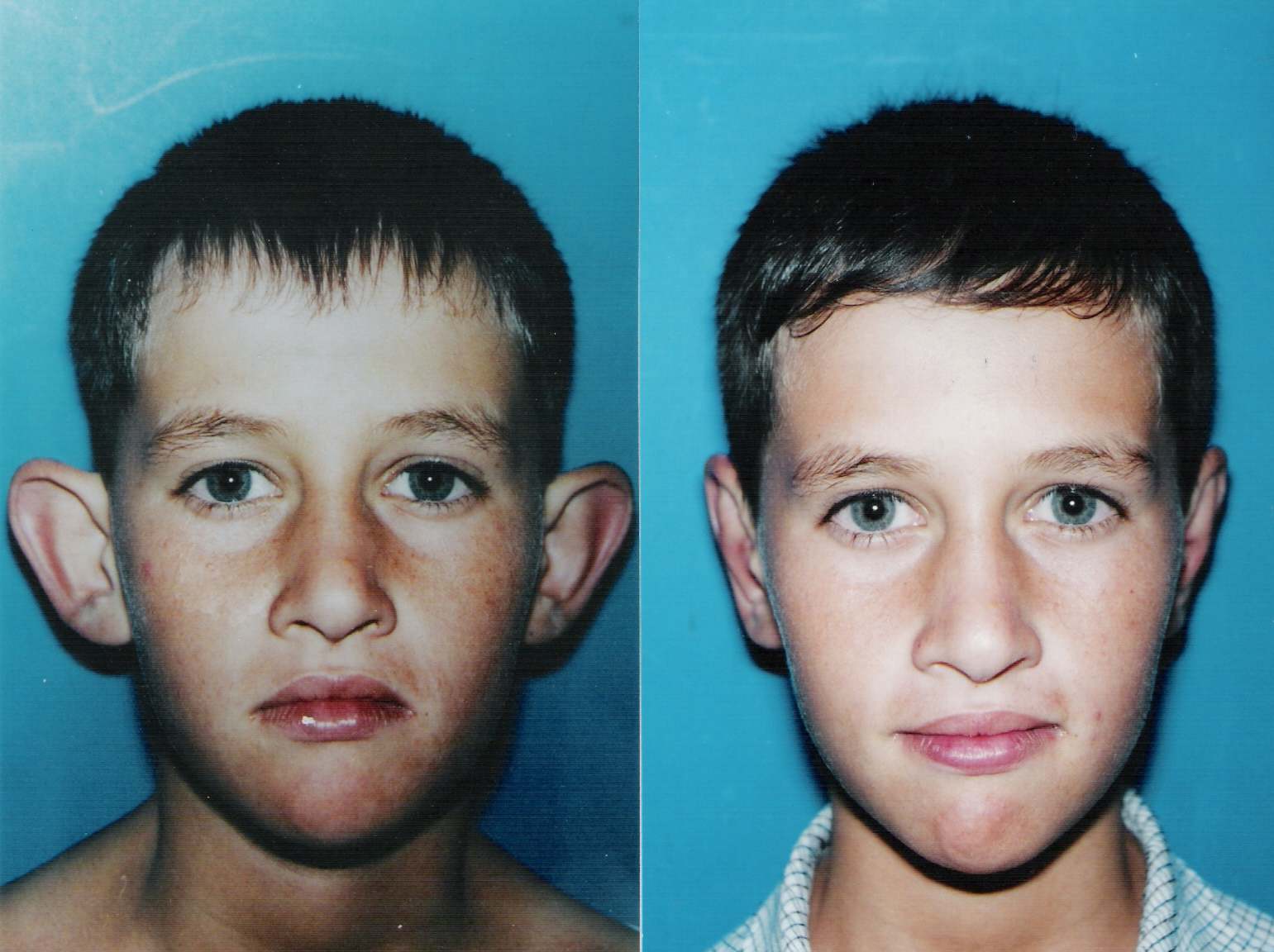
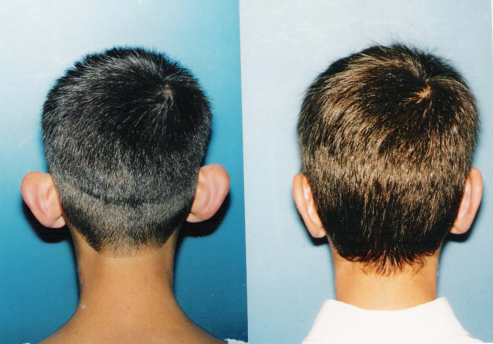
Fig. 7
View Synopsis (.doc format, 448.0 kb)