Sunday, October 8, 2006
10954
Scrotal Reconstruction Using Gracilis Muscle Flap in Fournier's Gangrene
Introduction and Purpose
Fournier's gangrene is a rare but fatal form of acute necrotizing fasciitis in the perineum and external genitalia in men. Aggressive and extensive debridement and use of the parenteral broad spectrum antimicrobial agents are the most important in the treatment. After control of infection and unavoidable loss of soft tissue, the major concern following Fournier's gangrene lies on the protection of the testicles and an adequate scrotal volume and appearance. This study presents our clinical experience, total of nine cases, in reconstruction of the penoscrotal region following Fournier's gangrene. The gracilis muscle flap and skin graft was used to regain the protection and an adequate appearance with adjunctive therapy of hyperbaric oxygen and exercise.
Materials and Methods
We experienced nine patients with Fournier's gangrene who underwent a soft tissue reconstruction using a piece of unilateral or bilateral gracilis muscle flap from 1999 to 2005. Ages ranged from 23 to 75 years (mean age, 48.3 years), and the mean hospital stay was 69.5 days (range, 46 to 96 days). The diagnosis was established from patient history and physical examination. Cultures initially performed showed a polymicrobial flora. All the patients underwent active early intervention such as extensive or sequential debridement, drug therapy using broad-spectrum antibiotics and hyperbaric oxygen therapy to stop the necrosis initially because the infection spreads quickly once the gangrene develops. Walking exercise was encouraged to promote wound healing and prevent flap atrophy. The scrotal reconstruction was carried out after carefully performing extensive debridement in an attempt to correct the deformity due to spermatic cord contracture and to prevent damaging the testes and cord. The gracilis muscle was elevated according to the conventional method. It was dissected from the origin to its insertion site to obtain the length of a flap sufficient to cover up to the penoscrotal junction through a subcutaneous tunnel. An attempt was made to prevent flap atrophy by not severing the obturator nerve distributing the gracilis muscle in the last seven patients who underwent the reconstruction.
Result
All the patients survived. Four patients had treatment history for diabetes mellitus and two patients had liver cirrhosis as underlying disease, but three patients had no other predisposing factors. There was no necrosis extending to the tunica vaginalis in any of these patients. The defect area was covered with bilateral gracilis muscle flaps in the six patients who underwent extensive debridement in the scrotum, external genitalia and abdominal wall. A unilateral gracilis muscle flap was used in three patients who underwent debridement limited to one scrotum and partial inguinal area. Urinary diversion was performed in all patients, and fecal diversion was done in one patient. The average period of hospitalization was 69.5 days (range, 46 to 96 days). The average period of hyperbaric oxygen therapy and exercise was 13 days (range: 0 to 23 days). No recurrence and special complications were noted during the time of hospitalization. Cosmetically satisfactory results were observed in all patients.
Conclusion
Fournier's gangrene was actively treated by prompt extensive debridement, antibiotics therapy, and hyperbaric oxygen therapy. The soft tissue defect in the scrotum, perineum and anterior abdominal wall as a result of the debridement were reconstructed using gracilis muscle flap transfer and skin graft. Functionally and cosmetically satisfactory outcomes with appropriate scrotal volume were obtained.
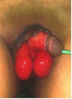
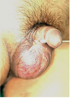
Fig 1. A 39-year old man. (Left) Before reconstruction, the denuded testicles with granulation tissue was noted. (Right) Appearance one month after surgery.
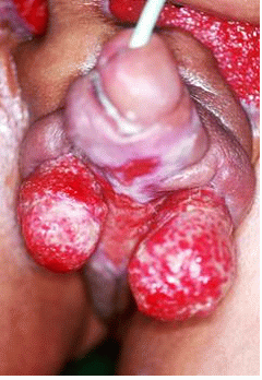
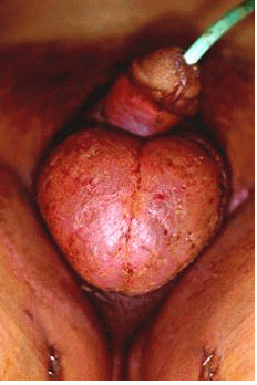
Fig 2. A 68-year old man. (Left) The soft tissue defect was noted on the scrotum and lower abdomen before the reconstruction. (Right) Appearance one month after surgery.
View Synopsis (.doc format, 421.0 kb)


