
Sunday, October 8, 2006 - 1:15 PM
11171
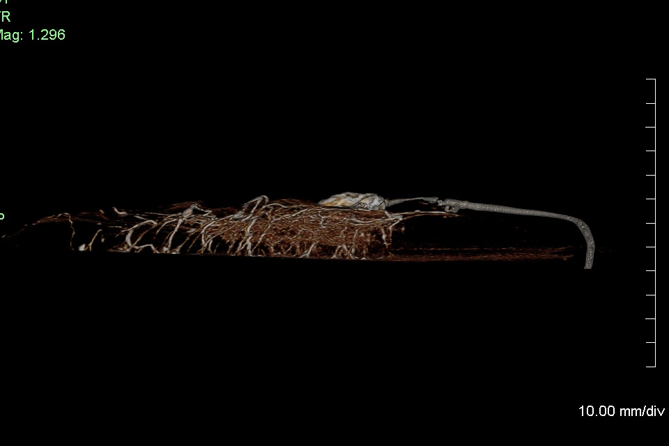
Four Dimensional Analysis of the Vascular Architecture and Perfusion of the Anterolateral Thigh Flap
Introduction: The vast majority of ALT flap anatomical vascular studies in the past have utilized lead oxide injections followed by 2-dimension radiography to determine vascular territories. Although lead oxide treated specimens provide excellent images, limitations of this methodology are well known. In contrast three-dimension radiography can provide not only qualitative data on vascular anatomy, but also information on the direction and location of blood flow within each layer of a perforator flap. Three dimensional anatomy was defined as the appraisal of the perforator vasculature in the sagittal, coronal and transverse views with an evaluation of the direction of blood flow through the perforator vascular tree, as seen in CT-angiography. Indeed to date, no studies have examined three and four dimensional vascular anatomies of the anterolateral thigh flap
Purpose: The goals of this study were two fold. First, to assess the static and dynamic vascular anatomy and branching pattern of the ALT perforator unit within the flap. Secondly, to establish a new comprehensive system of classifying the vascular branching patterns of the ALT perforator complex and correlate this with clinical applications.
Materials and Methods: Ten fresh cadaver ALT flaps were dissected suprafascially, based on the largest perforator originating from descending branch of the lateral femoral cutaneous artery. We then performed dynamic and static CT scans of all ALT flaps using a GE Light Speed 16 slice scanner. For dynamic scans, a Harvard pump was used to introduce 5 ml of iodinated contrast agent (Hypaque, Amersham Health) over a 10 min period. During the injection, helical scans were repeated at intervals to volume image the time evolution of flap vascularity. To minimize the volume scan time, the gantry was tilted to 30 degrees, and flaps were placed on a jig with a table angled to the gantry plane. Using a 0.5 s rotation time, 10 mm collimation (for 0.625 mm slices), and a 0.938 pitch setting, a 4 mm thick flap could be helically scanned in about 3 s. A similar geometry was used for static scans using lead oxide (Aldrich Chemical Co., Milwaukee, Wis.) or iodinated contrast agents, however, axial scanning at a 1.25 mm collimator setting (for 0.625 mm slices) was used to reduce artifacts. Scans were performed at 80 kVp when using iodinated contrast agent to optimize contrast, and 120 kVp was used for lead oxide contrast to minimize beam hardening artifacts (typically, scans were performed at ~ 200 mA). To allow optimal resolution reconstructions, raw scan data was saved to permit retrospective reconstruction in regions of interest.
For radiographs, of skin flaps, 5 ml of lead oxide (Aldrich Chemical Co., Milwaukee, Wis.) was injected Digital images were generated for each flap using computed radiography imaging. Each flap was placed directly on a computed radiography image plate cassette and radiographed at 80 kVp
Results: The main ALT perforator originated from the descending branch of the lateral femoral cutaneous artery in all flap dissections and nine of ten perforators were of the musculocutaneous variety. The ALT perforator unit vascular branching pattern was found to be highly variable and condensed throughout all layers of the flap with numerous vertical, oblique and horizontal vascular interconnections. Vascular communications between the fascial, adipose and dermal layers of the flap were observed up to the periphery of the flap in all cases and were maximized within a 5 cm radius of the perforator entry within the flap. Direct cutaneous, adipose and fascial perforator branches were observed in all Dynamic CT scan studies using iodinated contrast agent.
Conclusion: The ALT perforator unit branching pattern consists of a highly condensed network of direct and indirect branches linking the fascial, adipose and cutaneous components of the flap. This in turn provides additional insight in the possibility of safely harvesting large multi-component ALT flaps based on a single perforator.
View Synopsis (.doc format, 431.0 kb)
See more of Hand, Upper Extremity/Microsurgery
Back to 2006am Complete Scientific Program