Sunday, October 8, 2006
11216
Contour ThreadLift: Documenting Efficacy in 21 Patients
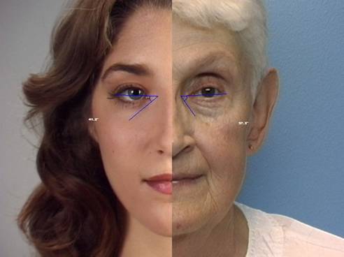
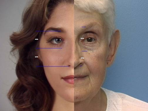
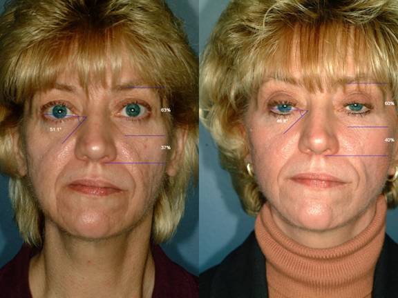
Pupose: To document whether Contour ThreadLift show improvement 6 months after surgery. Materials & Methods: Based on analysis of People magazine's beautiful people by decade, two measures were found to confirm “attractiveness.” These measures documented the perceived orbit as a percent of the midface and the angle formed by an intercanthal line and the tear trough shadow (tear angle). Ideally, the orbit should approach 50% of the midface, and /or the tear angle will approach 200. 21 Contour ThreadLift patients treated using the same deployment/tissue contouring technique were analyzed pre-operatively and 6 months post-operatively. Of the patients analyzed, 15 were ThreadLift only. Six patients had ThreadLift combined with another facial procedure, such as short scar facelift. All patients analyzed were treated with the CT-200 suture. Only changes in midface results were analyzed. Changes in jowls were not assessed. Results Summary: Of 21 patients, only one patient did not demonstrate measured improvement in the midface proportions. This patient's treatment goal of reducing jowls was met. Of the remaining 20 patients, all patients experienced improvements in the tear angle. Improvement varied from 0.20 to 12.60. Five patients experienced improvements of 100 or more. Thirteen of 21 patients experienced reductions in the orbital proportion of the midface, with reductions of up to 8% observed. In general, patients experiencing the greatest change in orbital proportion experienced less change in tear angle change. With only 1 exception, patients with combined procedures did not see significant changes in midface proportions or tear angle. Conclusions: Clinical improvement following Contour ThreadLift has been be documented. Prospective measurements can be used as a treatment planning tool, allowing management of both patient and surgeon treatment expectations. Following these patients, alterations in treatment technique have been implemented. Long term follow-up is not yet ready, though this same methodology will be used to assess both long term success, as well as assessment of treatment methods.
Fig 1: Demonstration of Tear Angle: Older vs Young

Fig.2: Demonstration of Midface ratios: Older vs Young

Fig. 3: Demonstration of Clinical Results
 Fig.4: Clinical planning: Study vs New deployment with pre-contoured soft tissue

