
Sunday, October 28, 2007 - 2:30 PM
12828
Four Dimensional Arterial and Venous Anatomies of the Deep Inferior Epigastric Perforator Flap
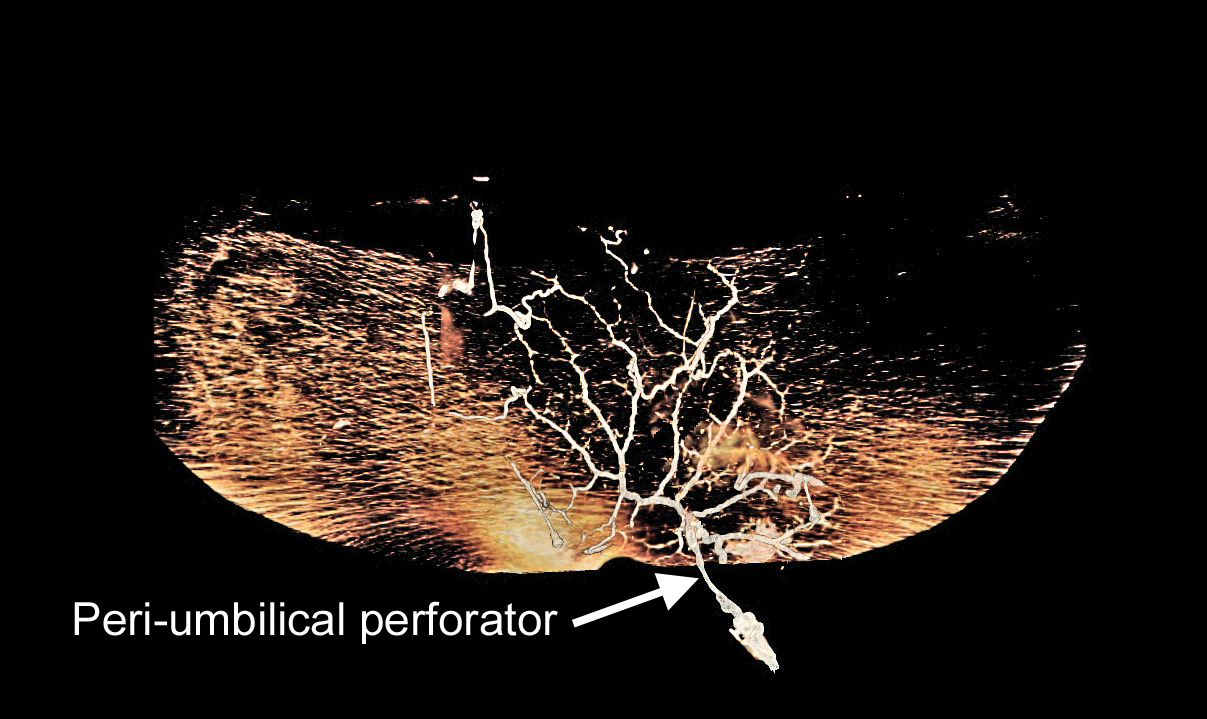
Background: In this study we investigate the vascular anatomy of the deep inferior epigastric artery perforator (DIEP) flap three- and four-dimensional computed tomographic (CT) angiography and venography.
Methods: Ten DIEP flaps harvested from fresh cadavers were included in the study. Dynamic and static computed CT angiography and venography using iodinated contrast medium were performed following cannulation of the largest medial and lateral row perforator bilaterally and the accompanying vena comitans. The perforators on one side of flap were scanned first, with washout of the contrast medium between scans, to compare perfusion when a medial or lateral row perforator was used. The direction of the valves within the venous system was confirmed by microdissection.
Results: The use of a lateral row perforator, in contrast to a medial row perforator, did not result in perfusion of zone IV. Perforators from the lateral row rely on recurrent flow through the subdermal plexus to perfuse the medial row perforators. In contrast, medial row perforators are connected across the midline by large diameter linking vessels at the level of the subdermal plexus. Histology confirmed that the medial row perforators have a thicker smooth muscle wall than the lateral row perforators in all cases. There is a superficial and deep venous drainage system, connected by venae commitantes, with dominance of the superficial inferior epigastric vein. Few branches cross the midline.
Conclusions: This study demonstrates that the medial row perforators are anatomically different to the lateral row perforators and should be selected if perfusion of zone IV is required. The poorly developed venous system across the midline may explain the frequent observation of venous compromise in the DIEP flap.
View Synopsis (.doc format, 1067.0 kb)
See more of Research and Award/Best Papers
Back to 2007am Complete Scientific Program