
Sunday, October 28, 2007 - 1:35 PM
13151
Bilateral Alar Reconstruction with Partially Split Septal Cartilage Graft –“Palm Tree Technique”- a 10-Year Experience with 56 Patients
Bilateral alar reconstruction with partially split septal cartilage graft –“Palm tree technique”- a 10-year experience with 56 patients.
Over-resection of the alar cartilages is a common problem faced by the plastic surgeon. A great variety of techniques have been proposed to reconstruct these structures, using cartilage grafts mounted and stitched in various manners. In patients with thin skin, though, any irregularity or distortion usually became visible postoperatively. Ishida and colleagues presented a molding technique to reproduce both alar cartilages in one piece, without stitches. This graft has the advantage of not having sharp edges or stitches that may distort or create irregularities to the nose. The aim of this study is to show a 10-year experience using this technique.
From January 1996 to July 2006, 56 patients were operated using the technique. A septal cartilage was harvested, leaving a 5-mm L-shaped strut. The graft is cut in an “L” shape with an approximate 135-degree angle (figure 1). The longer leg is split from its edge with a no.15 blade, preferably under magnification (figure 2). The splitting in performed until the transition with the shorter leg (figure 3). The graft is placed with its nonsplit portion fixed at the remnants of the medial crura (figure 4). This portion of the graft may work as a strut if necessary. The patients had a follow-up of a minimum of 6 months and a maximum of 10 years.
All patients had undergone one to five previous rhinoplasties, and remnants of the alar cartilages were not large enough to rebuild the cartilaginous structure. There was an improvement of the projection and three-dimensional conformation of the tip in all patients (figures 5 and 6). No postoperative irregularities or asymmetry were observed. None of the patients required re-operation in this series.
The splitting of the septum results in two leaves with curvature, thickness and elasticity very similar to the alar cartilages. As it is a one-piece graft, there is no need for stitches, and curvature of the domus is formed by the natural bending provoked by the splitting. This method reduces the chances of distortions, asymmetries and irregularities. Although this technique may result in a very compatible graft for bilateral alar reconstruction, it is a quite difficult maneuver to perform, especially with thin septal cartilages.
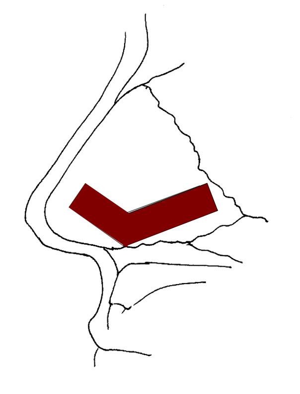
Figure 1 - Septal cartilage with demarcation of the graft to be harvest, in red.
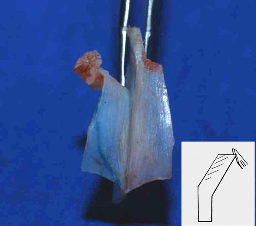
Figure 2- Septal cartilage with its cranial portion split by a blade no 15.
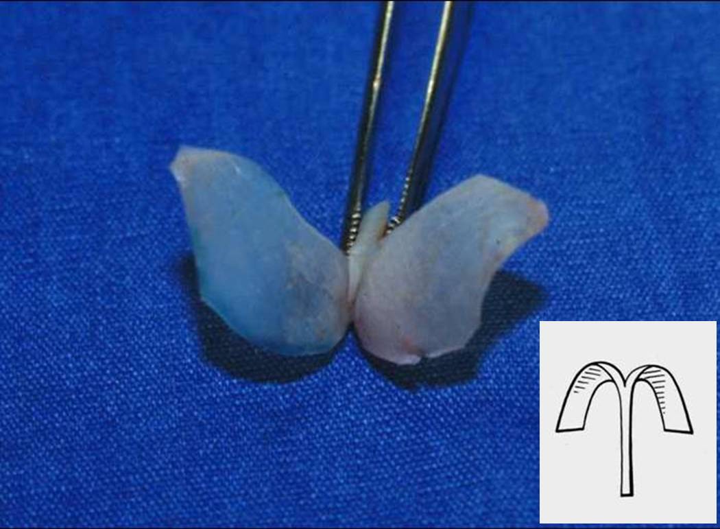
Figure 3 – Final appearance of the septal graft after molding.
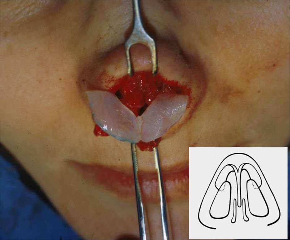
Figure 4- Fixation of the septal graft replacing both alar cartilages.

Figure 5- Patient with collapse of the alar cartilages secondary to a previous over-resection. On left side, preoperative pictures. On the right side, a 3-year postoperative view.
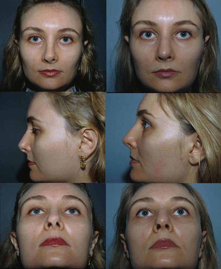
Figure 6- Patient with asymmetry of the alar cartilages visible due to a thin skin. On left side, preoperative pictures. On the right side, a 1-year postoperative view.
View Synopsis (.doc format, 417.0 kb)