Tuesday, November 4, 2008
14248
An Anatomical Study of the Superficial Peroneal Nerve Accessory Artery and Its Perforators, and Clinical Application of Superficial Peroneal Nerve Accessory Artery Perforator Flaps
An Anatomical Study of the Superficial Peroneal Nerve Accessory Artery and Its Perforators, and Clinical Application of Superficial Peroneal Nerve Accessory Artery Perforator Flaps
Background: In the 1990s, skin island flaps supplied by the vascular axis of sensitive superficial nerves, like the sural and saphenous nerves, were introduced. Flaps supplied by the superficial peroneal nerve accessory artery (SPNAA), however, are still not commonly used. The aim of this study is to understand the anatomical structure of the SPNAA and its perforators, and to utilize SPNAA perforator flaps in the clinic.
Methods: We dissected sixteen cadavers and assessed the location and number of the SPNAA, its perforators, and the septocutaneous perforators originating from the anterior tibial artery. The largest diameter of the SPNAA was also measured.
A SPNAA perforator flap was applied to thirteen patients, the free flap was applied to twelve patients, and the pedicled flap was applied to one patient.
Results: The origin of the SPNAA was 4 to 8 cm (average 5.5 cm) away from the fibular head. The SPNAA was 7 to 16 cm in length (average 12.33 cm), originating from the superior lateral peroneal artery and gradually disappearing between 15 and 22 cm (average 17.06 cm) from the fibular head. The largest diameter of the SPNAA was between 0.6 and 1.2 mm (average 0.85 mm). The number of perforators in SPNAA examined ranged from zero to eight (average 4.5). Of these, the number of septocutaneous perforators ranged from zero to six (average 3.19), and the number of musculocutaneous perforators ranged from zero to three (average 1.31).
The size of the flap ranged from 3.5 x 6 cm to 9 x 12 cm (mean 65.5 cm2). The complete follow-up period ranged from 6 to 24 months (mean 9 months). Although one flap was lost due to arterial thrombosis, the procedure was successful in the remaining twelve patients. The flap was very thin, comparable in thickness to the recipient site (i.e. foot, ankle, pretibia, knee or hand), and thus, aesthetically appealing.
Conclusion: SPNAA perforator flaps could be an excellent alternative to perforator flaps that use the lower leg as a donor site.
Legends
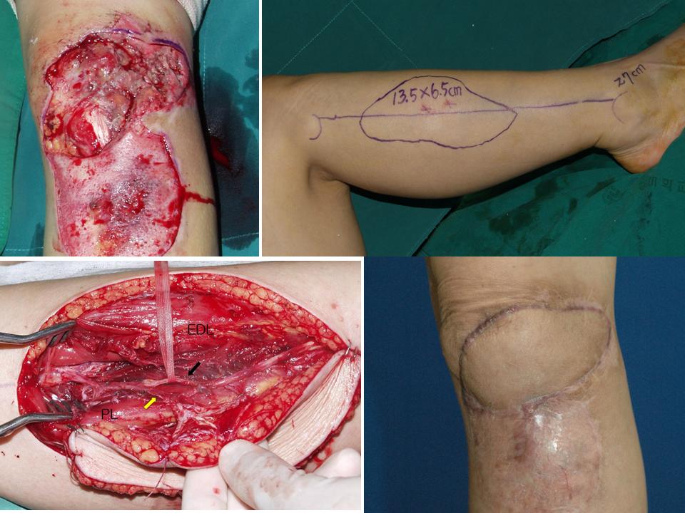
Fig. 1. (Above, left) Preoperative view. (Above, right) Preoperative design of the SPNAA perforator flap. (Below, left) The septocutaneous perforator of the SPNAA (yellow arrow) is the pedicle of this flap. Black arrow indicates the SPN. EDL: extensor digitorum longus. PL: Peroneus longus. (Below, right) Post-operative observation six months after surgery.

