Friday, October 31, 2008
14520
A NEW Treatment APPROACH for Sagittal Synostosis Surgery and Its Preliminary Results
Purpose: Various techniques have been performed for the correction of sagittal synostosis. The goal of modern craniofacial surgery is to achieve calvarial remodeling with minimally invasive surgical interventions. The aim of this study is to describe a new surgical technique for the treatment of sagittal synostosis and evaluate its results by assessing cranial morphologic changes over time.
Material and Methods: From years 2003 to 2006, 18 patients with nonsyndromic single suture sagittal synostosis have been operated with this new surgical technique. This technique is a simple cranioplasty which includes two thin bone strips craniectomy nearby the sagittal sinus and long parietal barrel-stave osteotomies. Anterior border of these strips is coronal suture, posterior border is lamdoid suture. Strips are located in 2 cm distance to sagittal suture and the width of these strips are 2 cm. Strips are fixed using resorbable systems and placed to a more lateral position than their original position (Fig. 1). Cranial lateral expansion is achieved due to pressure of the skin flap and elasticity of resorbable fixation system.
 |
Figure 1: The schematic presentation of the operational sequence.
3D Computed tomography, the cephalic index and the axial width ratio measures are obtained preoperatively and postoperatively at 6 and 12 months.
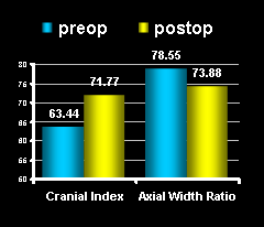
Results: The patient population consisted of 15 boys and 3 girls. The surgical age changed from 5 to 14 months (mean, 7.5 months), mean operation time was 120 minutes, ICU stay was 24 hours, hospital stay was 5 days and mean follow-up was 22 months (6-48). There were no major complications, BOS leakage, infections, mortality or the need for repeated remodeling procedure. The Cephalic Index changed from a mean preoperative value of 63.44% to a postoperative mean value of 71.77% (P = 0.008). The mean Axial Width Ratio changed from a preoperative 78.55% to 73.8% (P = 0.007). (Fig.<2)
\s 
Fig 2: Pre and postoperative cranial measurements.
Conclusions: This new technique can be used to allow for immediate lateral expansion. Improvement of cranial shape was obtained in early postoperative period for this lateral expansion. Although a few cranial indices have been found to be out of normal range, the difference between preoperative and postoperative cranial measures is statistically significant. This new procedure can be considered as an effective and safe surgical approach for sagittal synostosis treatment, evidenced by minimal morbidity, favorable cranial morphological changes and absence of mortality.
