
Friday, October 31, 2008
14536
Osseous regeneration of the free fibula flap used for mandibula reconstruction resembling a firm mass
AIM:
To discuss a case with bony regeneration resembling a firm mass after reconstruction of the mandibula with free fibula osteocutaneous flap
CASE REPORT:
Ten year old boy underwent surgery for mandibula reconstruction with osteo-cutaneous free fibula flap following a gun shot injury (Figure 1)
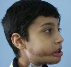
Figure 1
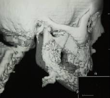
One year after a successful reconstruction, he complained about a firm mass on the angle of the mandibula at the reconstruction site. In 3-D tomography bony projection with 900 angle, 2x1x0,5 cm in size was seen at the posterior osteo-fibular junction (Figure 2)
 .
.
Figure 2
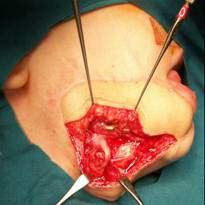
The bony segment was excised and the bone was burred (Figure 3,4).
 젨젨젨젨젨젨젨젨젨젨젨젨젨젨젨
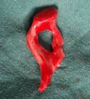
Figure 3젨젨젨젨젨젨젨젨젨젨젨젨젨젨젨젨젨젨젨젨젨젨젨젨젨젨젨젨젨젨젨젨젨젨 Figure 4
Histological examinations showed bony regeneration. Four months later he admitted with the same bony projection and he underwent re-excision and with a burr periosteum of the fibula flap was degenerated around the bony segment. The histological specimens corrected the bony regeneration
DISCUSSION:
Reconstruction of the mandibula with a free fibula flap is a common procedure. According to most head-neck surgeons because of the growth capacity, free fibula flap is the first choice in the reconstruction of critical sized mandibula defects in children. Bony projections are rare complications resulting from the vascularized periosteum that separated from the bone during the reconstruction. These bony projections may be very important in tumoral reconstructions by resembling a firm mass. Although it is very rare, one must keep this complication in mind. In the treatment, degeneration of the vascularized periosteum around the bony segment is essential for complete recovery.