Friday, February 1, 2008
13815
Surgical Treatment of Severe Graves' Dermopathy; A Case Report and Review of Literature
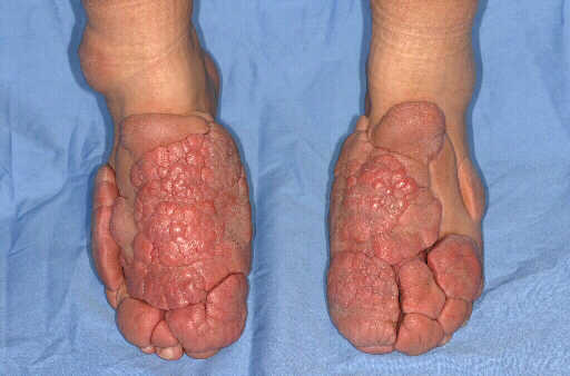
Purpose statement: Severe form of Graves' dermopathy is a rare manifestation of Graves' disease and provides a unique therapeutic challenge; since it's unresponsive to traditional medical therapy. Here, we present a case of severe Graves' dermopathy involving the dorsum of the foot treated surgically. Materials and Methods: A pleasant 36 year old woman presented with a 12 year history of multiple nodules and plaques of the dorsum of her feet and toes. She was diagnosed with Graves' disease 14 years ago and was treated with radioactive iodine. Eighteen months after her diagnosis she developed Graves' ophthalmopathy which was progressive and required orbital decompression. About two years after her diagnosis she developed right sided pretibial myxedema which involved the dorsum of her foot as well. She was treated with compressive therapy as well as steroids, however the lesions progressed. The lesions eventually developed into multiple large tuberous plaques which covered the dorsum of her feet as well as her toes. This progression was treated with subcutaneous somatostatin injections for a duration of one year. During this treatment there was some regression of the nodules, but eventually the lesions began to grow again. On presentation the patient had multiple large fungating indurated plaques; which had developed additional papulo-nodular lesions over them, giving a cobblestone appearance (Fig 1). These plaques were separated by islands of normal skin, which was completely covered by the overlying plaques, making it prone to maceration. The cobblestone skin atop the plaques would frequently ulcerate and bleed. Due to the size of the plaques which were 3 cm at greatest height, the patient was unable to wear most shoes and was developing difficulty walking. An MRI of the lesions showed: marked lobulated and nodular cutaneous soft tissue thickening limited to the skin, without evidence of extension into subcutaneous fat or tendons. Surgical therapy consisted of resection of lesions on the entire dorsum of the right foot along with the toes and a lateral maleolar lesion, followed by split thickness skin grafting. The resection was done to the level of the subcutaneous fat. Pathology results showed skin with extensive dermal mucinosis, consistent with myxedema. Results and Conclusions: The patient did quiet well post-operatively. She had complete take of her skin graft, and after a period of restricted activity is back to per-operative functional status. She is currently six months removed from her operation without any evidence of recurrence. Plan is for treatment of the other foot in the near future. This severe form of Graves' dermopathy affects less than 1% of patients with Graves' disease. There is sparse reports in the literature of the condition and the therapeutic challenge it presents. The reports come from the dermatology and endocrine literature, and give little in terms of successful treatment. Lessons from this case seem to suggest the excision with skin grafting is an excellent treatment option. In this short follow-up period there has been no recurrence. Recurrence however seems unlikely, since the affected dermis and dermal appendages were excised down to subcutaneous tissue. Longer follow-up is required to see if excision and skin grafting is a permanent solution.

