Monday, October 4, 2010 - 9:30 AM
17356
The Effect of Potassium Chloride in Rat Wound Healing
The effect of potassium chloride in rat wound healing
Introduction
Recent studies have found that the potassium channel is associated with wound healing and that wound healing can be promoted by regulating the potassium channels. Therefore, we hypothesized that regulating potassium chloride's concentration of the wound influence the activity of potassium channel, and have a good effect on wound healing. With experiments in the Sprague-Dawley rats, we planned to evaluate our hypothesis. In addition, we tried to inquire into the effect of wound healing in vitro.
Methods
Cell viability and cytoprotective effect of HaCaT (immortalized human keratinocyte cell line) and human neonatal dermal fibroblast (HNDF) in response to various concentrations of potassium chloride (10, 15 and 25 mM) were inspected. By western blot analysis, vascular endothelial growth factor (VEGF) and thrombospondin 1 (TSP1) of HaCaT which was stimulated by TNF-a (10 ng/ml) and pretreated by various concentrations of potassium chloride (10, 15 and 25 mM) were measured. By collagen assay, the changes of collagen production of HNDF under various concentrations of potassium chloride (10, 15 and 25 mM) were measured.
With rat experiments, 2x2 cm full thickness wounds were made on the back of 13 male Sprague-Dawley rats. 25 mM potassium chloride was applied to the experimental group and distilled water to the control group. Wound size was measured and western blot analysis was performed for VEGF, TSP1 and CD31.
Results
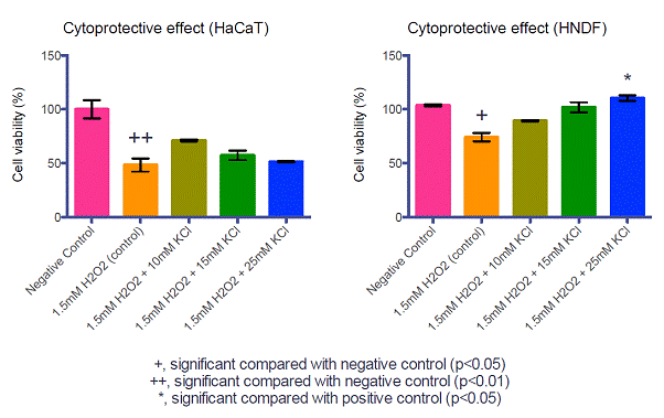
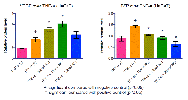
With various concentrations of potassium chloride, cell viability of experimental group had no difference with control group's (Fig 1). In cytoprotective effect study, cell viability was increased with potassium chloride treatment than H2O2 treated control group's (Fig 2). VEGF level in potassium chloride treated group was significantly higher than TNF-a only control group's, while TSP1 level was significantly lower (Fig 3). In collagen assay of HNDF, experimental group¢®¯s collagen production was greater than control group¢®¯s (Fig 4).
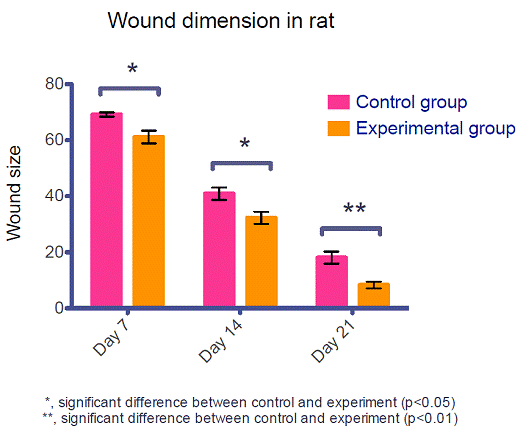
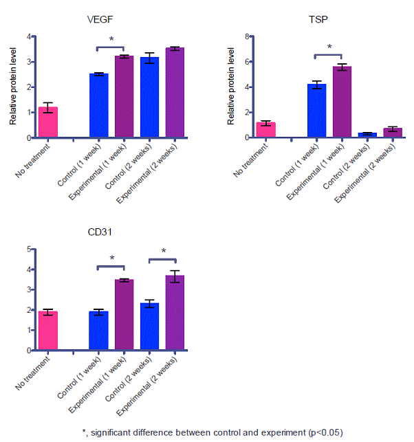
With rat experiments, potassium chloride treated experimental group healed faster than control (Fig 5). VEGF and CD31 were significantly increased in experimental group while TSP1 was decreased than control group (Fig. 6).
Conclusion
We observed the effect of potassium chloride in improvement of wound healing with cytoprotective effect, angiogenesis and collagen production in rat model.
Acknowledgements
This work was supported by the Korea Science and Engineering Foundation (KOSEF) grant funded by the Korea government (MOST) (No. R01-2007-000-20746-0).

Figure 1. Cell viability in HaCaT and HNDF under potassium chloride

Figure 2. Cytoprotective effect of potassium chloride in HaCaT and HNDF under H2O2

Figure 3. Protein levels of VEGF and TSP 1 in HaCaT cells (Western blot analysis)

Figure 4. Changes of collagen production in HNDF

Figure 5. Changes of wound size in rats experiment
 Figure 6. Protein levels of various cytokines in rats experiment (Western blot analysis)
See more of Research (5 mins)
Back to 2010am Complete Scientific Program
