
Room 2 (Henry B. Gonzalez Convention Center)
Sunday, November 3, 2002
8:00 AM - 4:00 PM
Room 2 (Henry B. Gonzalez Convention Center)
Monday, November 4, 2002
8:00 AM - 4:00 PM
Room 2 (Henry B. Gonzalez Convention Center)
Tuesday, November 5, 2002
8:00 AM - 4:00 PM
Room 2 (Henry B. Gonzalez Convention Center)
Wednesday, November 6, 2002
8:00 AM - 4:00 PM
541
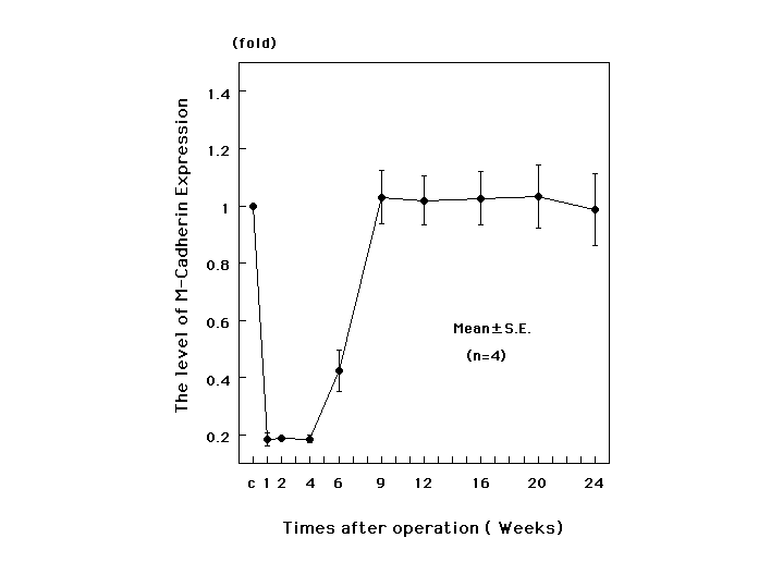
P17 - Detection of M-Cadherin Expression After Nerve Repair in a Rat Sciatic Nerve Model
It is well recognized that M-cadherin plays important roles in myogenesis. In the present study we investigated M-cadherin expression in skeletal muscle in denervation/reinnervation process. After rat sciatic nerve was resected by fine scissors, the nerve was sutured using 10-0 monofilament nylon suture. At various periods up to 24 weeks after the operation, the rats were killed by cervical dislocation, and the gastrocnemius muscle was removed. M-cadherin expression in the muscle was detected by Western blot analysis using anti-M-cadherin monoclonal antibody. The level of M-cadherin expression in the muscle was calculated as the relative amount to that found in normal control muscle. The level of M-cadherin expression was significantly low at 1,2,4,6 postoperative weeks, but the level returned to the control level on 9th postoperative week. The minimum level of M-cadherin was 0.184±0.01 fold at 1st postoperative week. The operated foot started to move at the 6th postoperative week, and the degree increased with time. Theseresults demonstrated that the level of M-cadherin expression in skeletal muscle decreased in denervation/reinnervation process. Moreover, it may be suggested that expressed M-cadherin plays important roles in the functional regeneration of the skeletal muscle.
View Synopsis (.doc format, 40.0 kb)
See more of Posters
Back to 2002 Complete Scientific Program
Back to 2002 Meeting home