
Room 2 (Henry B. Gonzalez Convention Center)
Sunday, November 3, 2002
8:00 AM - 4:00 PM
Room 2 (Henry B. Gonzalez Convention Center)
Monday, November 4, 2002
8:00 AM - 4:00 PM
Room 2 (Henry B. Gonzalez Convention Center)
Tuesday, November 5, 2002
8:00 AM - 4:00 PM
Room 2 (Henry B. Gonzalez Convention Center)
Wednesday, November 6, 2002
8:00 AM - 4:00 PM
982
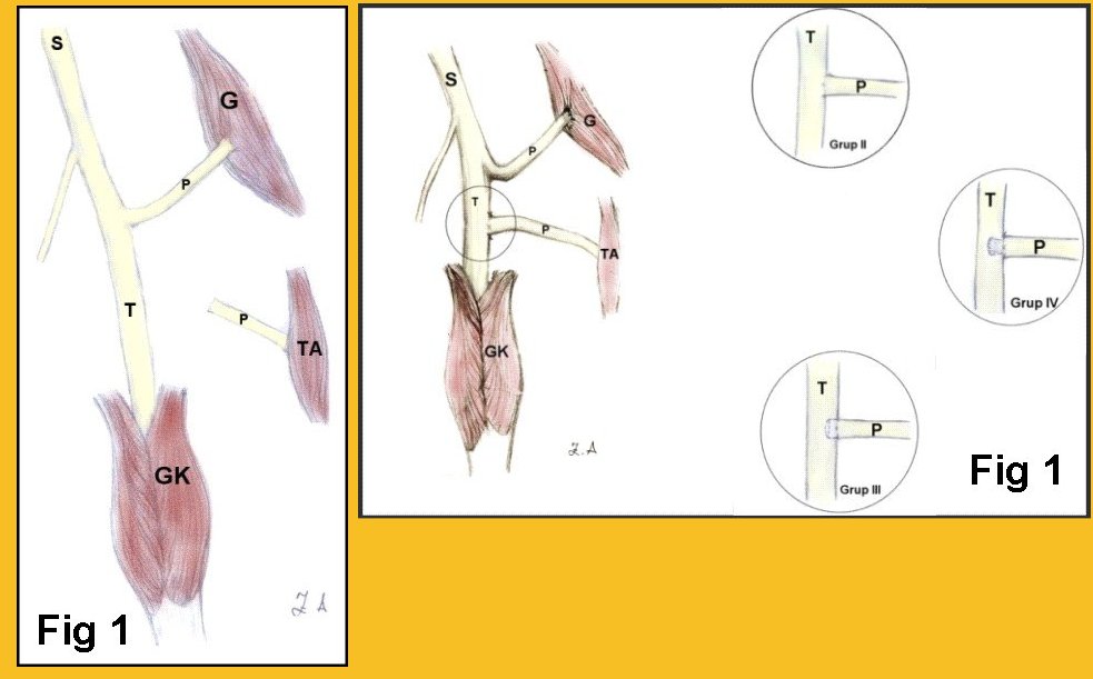
P54 - The Effect of Distal Epineural Resection of the Recipient Nerve on Axonal Regeneration in Terminolateral Neurroraphy
Introduction: When neither end-to-end neurroraphy nor nerve grafts are available for restoration of the nerve continuity following nerve damage, end-to-side neurroraphy is an alternative. Unfortunately, end-to-side neurroraphy is not successful yet to restore lost function enough as end-to-end neurroraphy. We aimed to find out the impact of distal epineural resection of the recipient nerve on axonal regeneration in end-to-side neurroraphy. Materials and Methods: Forty wistar male rats were included in this study as 4 groups. Under ketamine-HCl anesthesia, in the right hind-limb of the animals, peroneal nerve was transected at a level of about 1 cm above the bifurcation of the tibial nerve, and the proximal stump of the peroneal nerve was buried into gluteus maximus muscle in all groups. Distal peroneal nerve was not processed in-group I (Fig 1), was sutured to the epineurial window on tibial nerve by epineurial neurroraphy in-group II (Fig 2). Distal stump of the peroneal nerve was buried in tibial nerve for about 2 mm without any epineurial resection from peroneal stump in-group III (Fig 2), and with 2 mm epineurial excision in-group IV (Fig 2). Walking track analysis and electromyographic studies performed after 3 months, thereafter, animals were sacrificed and muscle weight measurement and muscle fiber analysis, histomorphometric nerve analysis including axon count, axon diameter and histological analysis were done. Statistical analysis was done with SPSS 10.0 software. Results: No functional loss in donor nerve or in donor muscle was determined. Healing was superior in-group IV. No statistically important difference was found out between groups II, III and IV but group I. Conclusion: End-to-side anastomosis supplies a degree of peripheral nerve regeneration without any evident donor nerve function loss. However, new studies must be done for improving regenerative capacity of the technique.
View Synopsis (.doc format, 436.0 kb)
See more of Posters
Back to 2002 Complete Scientific Program
Back to 2002 Meeting home