
Wednesday, October 13, 2004 - 7:10 AM
5710
Distally Based Lateral and Medial Leg Adipofascial Flaps: Be Cautious in Old and Diabetic Patients
Introduction: Defects around the ankle result from various etiologic factors, but repair of the diabetic ulcers requiring a flap represents a major concern for plastic surgeons dealing with lower extremity reconstruction. Because, the incidence of peripheral vascular disease in these patients is at least four times higher and there is a predilection for occlusive disease to involve primarily the tibial and peroneal arteries between the knee and the ankle, and coverage of the malleolus, Achilles tendon or heel has been specially troublesome. Lateral and medial leg adipofascial flaps distally based on perforators originating from the peroneal and posterior tibial arteries, respectively, were reported to be encouraging in reconstruction of these defects. Presented here is our clinical experience with these two flaps particularly emphasizing the complicated attempts in diabetic patients. Material and Methods: Seven patients (2 male and 5 female) having skin defects with bone or tendon exposure around the ankle were treated with lateral and medial leg adipofascial flaps. The lowermost perforators of the peroneal or posterior tibial artery were identified preoperatively with a hand-held Doppler and a straight incision, through skin only, was made proximal to this perforator down to the adipose tissue and long enough to allow tension-free positioning of the flap. The skin edges were dissected in the subdermal plane leaving only a thin layer of superficial adipose tissue on the skin flaps. With the skin flaps reflected, the adipofascial flap was than raised in the subfascial plane. The perforators to be retained in the base were located and the flap was then turned over to cover the defect so that the adipose tissue laid deep and the fascia became superficial followed by application of a split-thickness skin graft over the flap. Donor site was closed primarily. Results: The ages of the patients ranged from 25 to 80 years, and the size of the flaps ranged from 3 x 5 cm to 7 x 10 cm. Follow-up time varied from 1 to 11 months (Table). Four defects were reconstructed with lateral leg adipofascial flap and medial leg adipofascial flap was utilized in three. Two flaps (1 lateral and 1 medial) healed uneventfully. Partial or total graft loss and partial flap necrosis were observed in 5 patients, 4 of whom were diabetic (Figures 1 and 2). Debridement and second grafting (third in 2 cases) was performed in these diabetic patients to achieve a stable reconstruction. Conclusion: Leg adipofascial flaps with advantages including a wide arc of rotation, minimal donor site morbidity and an easy dissection without sacrifice of a major artery, offer a valuable option for repair of defects around the ankle in many cases. However, diabetic ulcers constitute a significant fraction of these lesions and adipofascial flaps should be used with caution in old and diabetic patients. Yet when performed, the probability of a second or third procedure should be taken, or skin grafting might perhaps be delayed until the viability of the flap is certain.

Table: Clinical data of the patients.
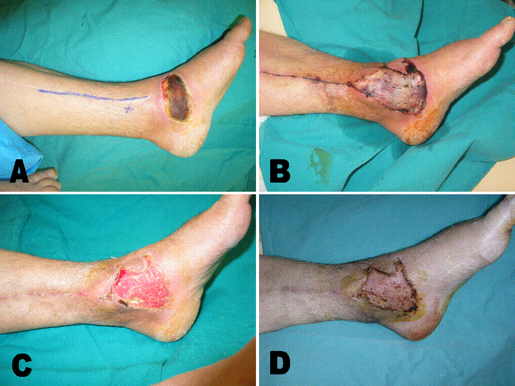 |
Figure 1 (Case 3): A; diabetic ulcer on the medial malleolus, skin incision and the lowermost perforator of the posterior tibial artery are marked. B; postoperative thirteenth day, skin graft did not take well. C; postoperative day 40, after debridement of the necrotic graft, granulation tissue covered the flap. D; one month after second grafting (75 days after the first operation), no problems were observed thereafter.
 |
ĀĀ
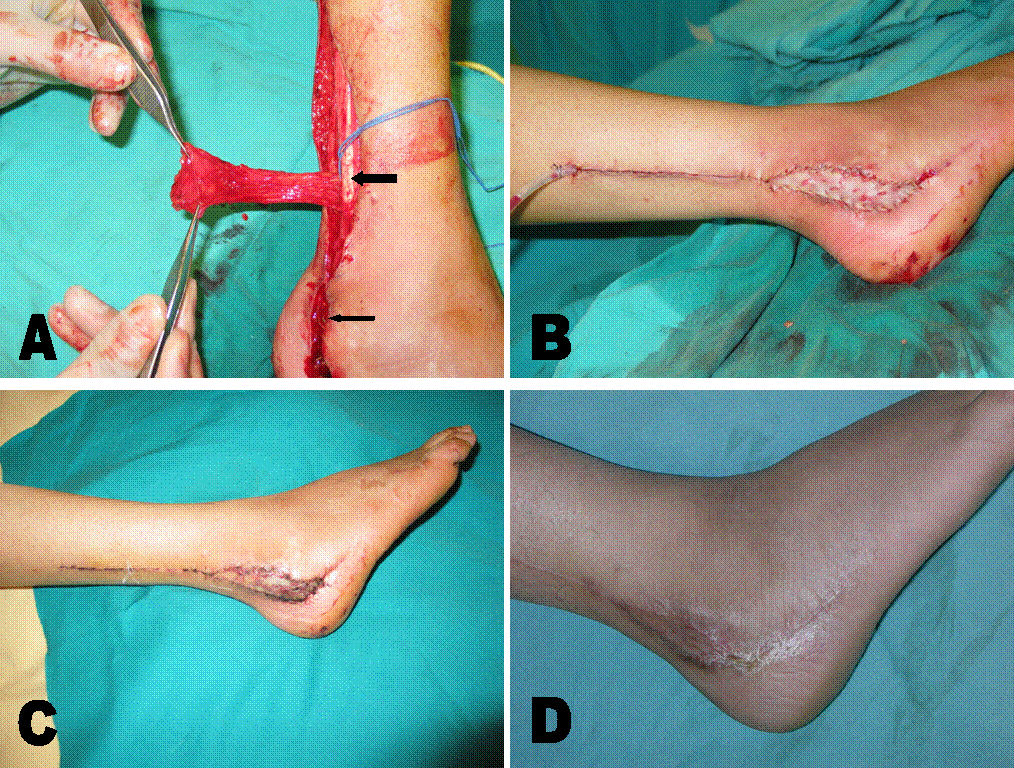
Figure 2 (Case 4): A; the flap is raised, lowermost perforator of the peroneal artery (thick arrow) and the exposed plate (thin arrow) are seen. B; the flap is inset and resurfaced with graft, donor site is closed. C; partial graft loss at the tip of the flap at postoperative day 5. D; five months after the procedure, the area of graft necrosis healed secondarily.
View Synopsis (.doc format, 89.0 kb)
See more of General Reconstruction
Back to 2004am Complete Scientific Program