
Wednesday, October 13, 2004 - 7:00 AM
5940
Dorsal Intercostal Perforator Flap: A New Option For Back Reconstruction
INTRODUCTION: The posterior intercostal arteries, by their many perforators, form the largest angiosome in the torso. Our comparative angiographic study between human and animals 1) revealed that the dorsal perforators of the posterior intercostal arteries constantly existed and they were large enough to supply their own vascular territories in the overlying skin. Clinically, the back is the source of the most useful surgical flaps such as the latissimus dorsi flap, the trapezius flap or the scapular flap. Those flaps, however, must be unfeasible if their main pedicles or muscles are spoiled in the previous surgeries around shoulder and axilla. On these anatomic bases and clinical demands, the dorsal intercostal perforator (DIP) flap was developed as a new island flap.
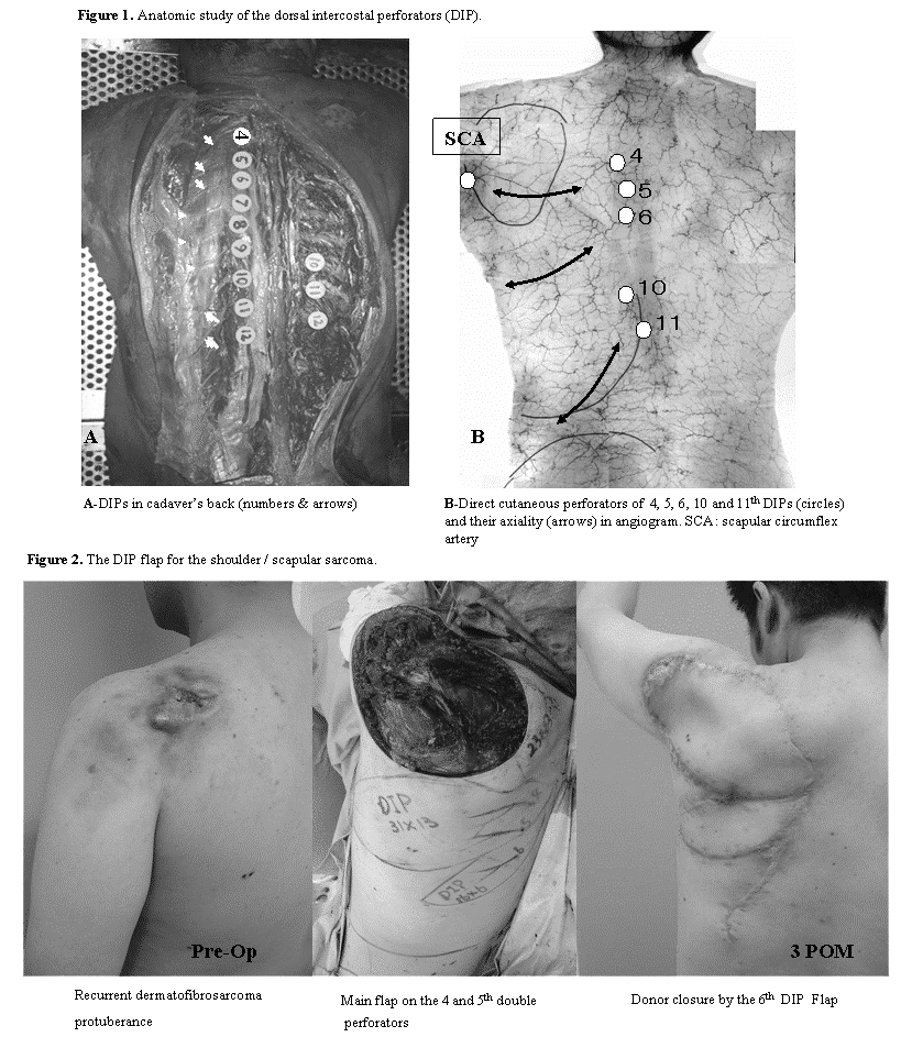
ANATOMY AND OPERATIVE TECHNIQUES: According to our anatomic study (Fig. 1), each of 4th to 12th posterior intercostal arteries constantly sent off the dorsal perforators. Those dorsal intercostal perforators (DIPs) were seemed to be the Åemedial' dorsal cutaneous branches of the posterior intercostal arteries 2). The DIPs from the 4th, 5th, 6th, 10th and 11th posterior intercostal arteries located theirselves by the upper or the lower margin of the latissimus dorsi muscle and were the direct cutaneous perforators penetrating the trapezius muscle or the lumbodorsal fascia. The locations of the DIPs in the skin were about 1 to 2 inches lateral to the spinous processes of the vertebrae and were clinically detectable by the doppler probe preoperatively. The 4th, 5th and 6th DIPs anastomosed with transverse branches of the scapular circumflex artery (SCA) and they could remarkably extend their vascular territories as far as to the mid-axillary line. The 10th and 11th DIPs ran parallel to the costal angle, showing more vertical axiality to reach iliac crest. The 7th, 8th and 9th DIPs, covered by the latissimus dorsi muscle, commonly were less dominant than other DIPs. The DIP flaps were elevated as fasciocutaneous island or pedicled flaps, preserving the latissimus and/or the trapezius muscles in the donor site. On the contrary, it was possible to harvest the DIP flap even if the underlying muscles or the SCA had been ablated. The dissection of the flap could easily and safely be carried out, starting from the distal end of the flap and following the under surface of the fasciae with attention to the musculocutaneous perforators. Each DIP, as a vascular pedicle, was noticeable, when the dissection was approaching to the dorsal midline. As the DIPs located segmentally, a couple of DIPs could be contained in one flap to extend the flap dimension.
CASES AND RESULTS: Ten DIP flaps were applied in nine cases for the reconstruction of upper back sarcomas (4 cases), an empyema, a vertebral pressure sore and the tissue defects of large flaps donor sites (4 cases). In eight cases, the muscles of latissimus dorsi or trapezius, or the SCA were spoiled in the previous surgeries. The maximum flap dimension was 31 x 13 cm in size containing double DIPs, which donor site was closed by another DIP flap (Fig. 2). All flaps showed stable postoperative blood circulation and almost survived completely only with a marginal necrosis in the largest flap. No functional loss owing to the DIP flap harvest was recognized in every case.
CONCLUSIONS: As depending on the segmental dorsal intercostal perforators, the DIP flap is always feasible only if the dorsal midline is preserved nevertheless of the previous surgeries. Based on the consistent multiple vascular pedicles with no need of dissecting the underlying muscles, the DIP flap is a new, less invasive option to be considered for the back reconstruction.
References
1. Taylor, G.I., and Minabe, T. The angiosomes of the mammals and other vertebrates. Plast. Reconstr. Surg. 89: 181, 1992.
2. Cormack, C. G., and Lamberty, B. G. H. The arterial anatomy of skin flaps, 2d Ed. London: Churchill-Livingstone, 1994.
View Synopsis (.doc format, 274.0 kb)
See more of General Reconstruction
Back to 2004am Complete Scientific Program